. 2006 Mar;26(5):1598-609.
doi: 10.1128/MCB.26.5.1598-1609.2006.

DNA damage during reoxygenation elicits a Chk2-dependent checkpoint response

Affiliations

DNA damage during reoxygenation elicits a Chk2-dependent checkpoint response

Rachel A Freiberg et al. Mol Cell Biol. 2006 Mar.

Abstract

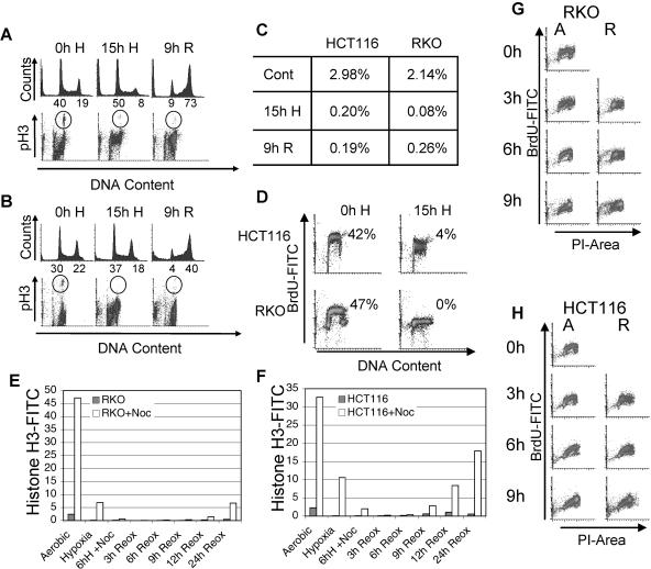
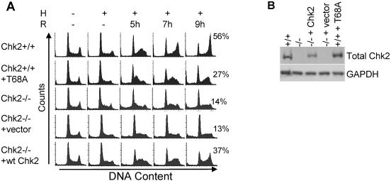
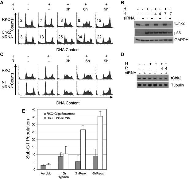
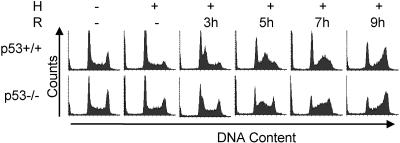
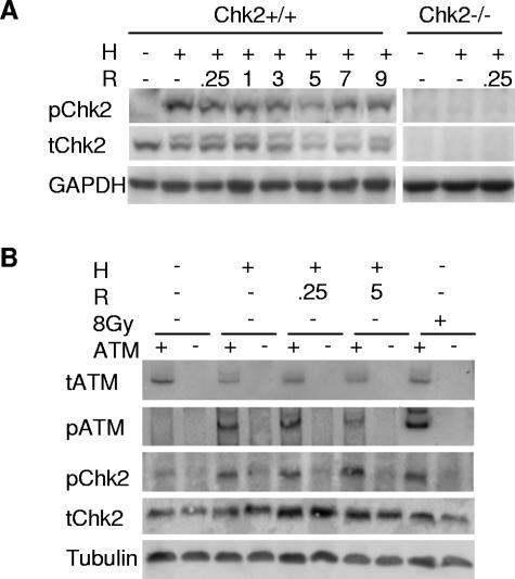
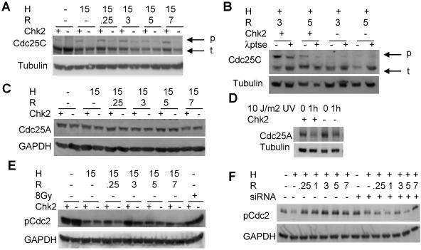
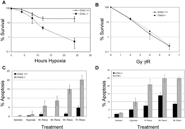
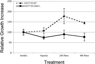
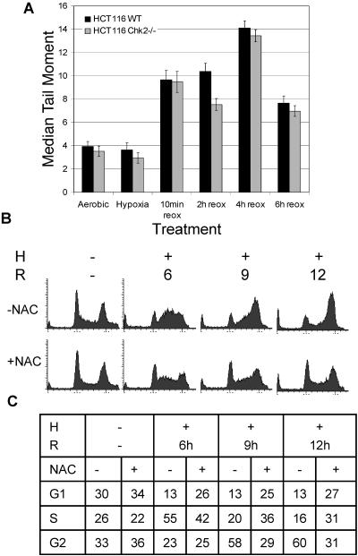
Due to the abnormal vasculature of solid tumors, tumor cell oxygenation can change rapidly with the opening and closing of blood vessels, leading to the activation of both hypoxic response pathways and oxidative stress pathways upon reoxygenation. Here, we report that ataxia telangiectasia mutated-dependent phosphorylation and activation of Chk2 occur in the absence of DNA damage during hypoxia and are maintained during reoxygenation in response to DNA damage. Our studies involving oxidative damage show that Chk2 is required for G2 arrest. Following exposure to both hypoxia and reoxygenation, Chk2-/- cells exhibit an attenuated G2 arrest, increased apoptosis, reduced clonogenic survival, and deficient phosphorylation of downstream targets. These studies indicate that the combination of hypoxia and reoxygenation results in a G2 checkpoint response that is dependent on the tumor suppressor Chk2 and that this checkpoint response is essential for tumor cell adaptation to changes that result from the cycling nature of hypoxia and reoxygenation found in solid tumors.


Figures

FIG. 1.
FIG. 1.
Reoxygenation-induced arrest in HCT116 and RKO cell lines. Colon carcinoma cells arrest in the G2 phase of the cell cycle following exposure to hypoxia and reoxygenation. FACS analysis of PI-stained DNA and FITC-labeled pH3 in HCT116 (A) or RKO (B) cells either untreated (0h H), exposed to 15 h of hypoxia (15h H), or exposed to 15 h hypoxia and 9 h of reoxygenation (9h R). The percentages of cells in G1 and G2 are indicated below the axes of the histograms. (C) Quantification of FITC-pH3 staining in panels A and B. (D) HCT116 and RKO cells undergo replication arrest during exposure to hypoxia. FACS analysis of BrdU incorporation in untreated cells (0h H) and those exposed to 15 h of hypoxia (H). RKO (E) and HCT116 (F) cells were subjected to hypoxia and reoxygenation (Reox). Cells were arrested in G2 by the addition of nocodazole during hypoxia or during reoxygenation and then analyzed by FACS for FITC-labeled phospho-histone H3 and DNA stained with propidium iodide. For hypoxic samples, nocodazole (Noc) was added immediately or after 6 h of hypoxia (6h H), while for reoxygenation samples, nocodazole was added immediately upon removal from hypoxia. Both RKO (G) and HCT116 (H) were exposed to 15 h of hypoxia and up to 9 h of reoxygenation. S-phase cells were labeled with BrdU for 1 hour between 2 and 3 hours of reoxygenation. For a control (0), untreated cells were also labeled with BrdU for 1 hour. FACS analysis of FITC-BrdU- and propidium iodide-labeled cells is shown. Hours of either aerobic conditions (A) or reoxygenation (R) are shown along the FITC-BrdU axes.
FIG. 2.
FIG. 2.
Reoxygenation-induced G2 arrest is dependent on the protein kinase Chk2. (A) Cells were untreated (−) or treated with 15 h of hypoxia (H), followed by indicated hours of reoxygenation (R). Cell types are HCT116 Chk2+/+, HCT116 Chk2+/+ stably expressing Chk2T68A, HCT116 Chk2−/−, HCT116 Chk2−/− stably expressing empty vector, and HCT116 Chk2−/− stably expressing Chk2+/+. Histogram profiles of DNA content were derived from FACS analysis from PI staining. The percentage of cells in the G2 phase of the cell cycle is shown for 9 h of reoxygenation next to each histogram. (B) Confirmation of stable integration of Chk2+/+-expressing vector in HCT116 Chk2−/− cells. Total protein (50 μg) was run and transferred to a polyvinylidene difluoride membrane and then probed with total Chk2 antibody and an antibody to GAPDH for loading control.
FIG. 3.
FIG. 3.
Reoxygenation-induced G2 arrest after Chk2 siRNA treatment is attenuated in the RKO colon carcinoma cell line. (A) Chk2 protein knockdown in RKO colon cancer cells using Chk2 siRNA. RKO cells treated with or without Chk2 siRNA were subjected to 15 h of hypoxia (H) followed by 3, 6, or 9 h of reoxygenation (R). Shown here are histograms generated by FACS analysis of PI-stained DNA. (B) Knockdown of Chk2 protein by siRNA. Immunoblots of protein extracts from cells treated with 15 h of hypoxia followed by reoxygenation were probed with antibody to total Chk2 (tChk2), total p53, and GAPDH. (C) G2 arrest induced by Chk2 siRNA treatment was specific. NT-siRNA oligonucleotide treatment had no effect on cell cycle response to hypoxia and reoxygenation. Histograms were generated by FACS analysis of PI-stained DNA. (D) Western blots on protein extracts from NT-siRNA-treated cells subjected to hypoxia and reoxygenation probed with antibodies to total Chk2 and α-tubulin. (E) Quantification of sub-G1 populations in RKO cells treated with or without Chk2 siRNA and exposed to hypoxia and reoxygenation.
FIG. 4.
FIG. 4.
G2 arrest is independent of p53 status. HCT116 wt and p53−/− cells were untreated (−) or treated with hypoxia (H) and reoxygenation (R). Shown here are histograms generated by FACS analysis of PI-stained DNA.
FIG. 5.
FIG. 5.
Chk2 phosphorylation is ATM dependent during hypoxia and reoxygenation. (A) HCT116 Chk2+/+ and Chk2−/− cells were treated with 15 h hypoxia (H) and reoxygenation (R) for the indicated hours. Protein lysates were subjected to Western blotting. Phospho-T68 Chk2 is shown in the upper panel, total Chk2 protein is shown in the middle panel, and GAPDH loading control is shown in the bottom panel. Lysates from HCT116 Chk2−/− cells failed to react with either Chk2 antibody (right panels). (B) Chk2 phosphorylation in a lymphoblastoid cell line treated with hypoxia and reoxygenation. The lymphoblastoid cell lines GM0536 (ATM+/+) and GM1526 (ATM−/−) were subjected to 15 h of hypoxia and various hours of reoxygenation. Western blots of protein lysates were probed with antibodies to phospho-S1981 ATM (pATM), total ATM (tATM), phospho-T68 Chk2 (pChk2), and α-tubulin (Tubulin).
FIG. 6.
FIG. 6.
Chk2 targets are phosphorylated following hypoxia and reoxygenation. (A) Cdc25C is phosphorylated during reoxygenation and is dependent on Chk2. HCT116 Chk2+/+ and Chk2−/− cells were treated with 15 h of hypoxia (H) and various times of reoxygenation (R). The top panel shows phosphorylated (p) forms migrating more slowly than total Cdc25C (t). An α-tubulin loading control is shown in the bottom panel. (B) Lambda protein phosphatase (λptse) treatment of protein extracts from HCT116 Chk2+/+ and Chk2−/− cells harvested after hypoxia and reoxygenation. (C) Cdc25A is not phosphorylated or degraded during hypoxia or reoxygenation treatment. HCT116 Chk2+/+ and Chk2−/− protein lysates from cells treated with 15 h of hypoxia and indicated times of reoxygenation were subjected to Western blotting and detection with total Cdc25A antibody, as shown in the top panel. GAPDH loading control is shown in the bottom panel. (D) Cdc25A is phosphorylated and degraded in response to UV stress. HCT116 Chk2+/+ and Chk2−/− cells were subjected to 10 J/m2 UV light. Cells were lysed after 1 h. Cdc25A was detected with total Cdc25A antibody after Western blotting, as shown in the top panel. α-Tubulin loading control is shown in the bottom panel. (E and F) Cdc2 phosphorylation during reoxygenation is Chk2 dependent. Protein lysates from HCT116 Chk2+/+ and Chk2−/− cells or from RKO cells treated with Chk2 siRNA were treated with 15 h hypoxia and indicated times of reoxygenation and were probed with phospho-specific Tyr15 Cdc2 (pCdc2) antibody after Western blotting, as shown in the upper panels. GAPDH loading control is shown in the lower panels.
FIG. 7.
FIG. 7.
Chk2−/− cells are sensitive to hypoxia and reoxygenation. (A) Colony-forming efficiency. Decreased survival of HCT116 Chk2−/− cells during reoxygenation is proportional to length of exposure to hypoxia. HCT116 Chk2+/+ and Chk2−/− cells were untreated or treated with hypoxia for 4, 8, 12, or 24 h and then incubated undisturbed for 2 weeks. Colonies were stained with crystal violet and counted. Results are expressed in log values. (B) Chk2+/+ and Chk2−/− cells are equally as sensitive to ionizing radiation. HCT116 Chk2+/+ and Chk2−/− cells were subjected to 0, 2, 4, 6, and 8 Gy gamma irradiation (γIR) and then allowed to grow undisturbed for 2 weeks. Colonies were stained with crystal violet and counted. Colony numbers are expressed in log values. (C) Increased apoptosis in Chk2−/− cells during reoxygenation. HCT116 Chk2+/+ and Chk2−/− cells were either untreated or placed in hypoxia for 15 h. Upon reoxygenation, cells were stained with Hoechst 33342 and PI. Nuclear morphology was assessed for each condition. Graphs are presented as the percentages of apoptotic cells in the total population. (D) Cells deficient in ATM are sensitive to reoxygenation. ATM+/+ (YZ3) or ATM−/− (pEBS) cells were treated with 15 h of hypoxia and then stained with Hoechst 33342 and PI at the indicated times. Results are reported as the percentages of apoptotic cells in the total population.
FIG. 8.
FIG. 8.
Chk2+/+ cells have a survival advantage during reoxygenation. HCT116 Chk2+/+ and Chk2−/− cells stably expressing EYFP and ECFP were mixed in equal numbers and plated onto slides and then either left untreated, treated with hypoxia overnight, or treated and then allowed to grow for 1 or 2 days under normal culture conditions. Blue and yellow cells were counted at each time point. Cell numbers relative to the starting population are plotted above.
FIG. 9.
FIG. 9.
Reoxygenation-dependent damage response is attenuated by ROS scavenger NAC. (A) The comet assay detects increasing damage during reoxygenation in HCT116 Chk2+/+ and Chk2−/− cells. Hypoxic samples were trypsinized and lysed under hypoxia conditions. Reoxygenation samples were harvested after indicated times. (B) HCT116 cells were treated with 10 mM NAC prior to hypoxia (H) and reoxygenation (R). After fixation, cell cycle distributions were assessed by FACS analysis of PI-stained DNA.

References

    1. Abraham, R. T. 2001. Cell cycle checkpoint signaling through the ATM and ATR kinases. Genes Dev. 15:2177-2196. - PubMed
    1. Adamson, A. W., D. I. Beardsley, W. J. Kim, Y. Gao, R. Baskaran, and K. D. Brown. 2005. Methylator-induced, mismatch repair-dependent G2 arrest is activated through Chk1 and Chk2. Mol. Biol. Cell 16:1513-1526. - PMC - PubMed
    1. Ahn, J. Y., J. K. Schwarz, H. Piwnica-Worms, and C. E. Canman. 2000. Threonine 68 phosphorylation by ataxia telangiectasia mutated is required for efficient activation of Chk2 in response to ionizing radiation. Cancer Res. 60:5934-5936. - PubMed
    1. Bakkenist, C. J., and M. B. Kastan. 2003. DNA damage activates ATM through intermolecular autophosphorylation and dimer dissociation. Nature 421:499-506. - PubMed
    1. Bartek, J., J. Falck, and J. Lukas. 2001. CHK2 kinase-a busy messenger. Nat Rev. Mol. Cell Biol. 2:877-886. - PubMed

Publication types

MeSH terms