Combination therapy of disseminated coccidioidomycosis with caspofungin and fluconazole
- PMID: 16480497
- PMCID: PMC1386678
- DOI: 10.1186/1471-2334-6-26
Combination therapy of disseminated coccidioidomycosis with caspofungin and fluconazole
Abstract
Background: The current recommended therapy for diffuse coccidioidal pneumonia involves initial treatment with amphotericin B deoxycholate or high-dose fluconazole, followed by an azole after clinical improvement. Amphotericin B is more frequently used as initial therapy if the patient's deterioration is rapid.
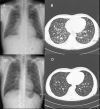
Case presentation: A 31-year-old Korean male with coccidioidomycosis presented to the hospital with miliary infiltrates on chest X-ray (CXR) and skin rash on the face and trunk. Initially, the patient did not respond to amphotericin B deoxycholate therapy. However, following caspofungin and fluconazole combination therapy, the patient showed favourable radiological, serological, and clinical response.
Conclusion: This appears to be the first case of diffuse coccidioidal pneumonia with skin involvement in an immunocompetent patient who was treated successfully with caspofungin and fluconazole. Combination therapy with caspofungin and fluconazole may, therefore, be an alternative treatment for diffuse coccidioidal pneumonia that does not respond to amphotericin B deoxycholate therapy.
Figures

Similar articles
-
Therapeutic efficacy of caspofungin alone and in combination with amphotericin B deoxycholate for coccidioidomycosis in a mouse model.J Antimicrob Chemother. 2007 Dec;60(6):1341-6. doi: 10.1093/jac/dkm383. Epub 2007 Oct 12. J Antimicrob Chemother. 2007. PMID: 17934204
-
Combination of caspofungin and an azole or an amphotericin B formulation in invasive fungal infections.J Infect. 2006 Jan;52(1):67-74. doi: 10.1016/j.jinf.2005.01.006. J Infect. 2006. PMID: 16368463
-
Coccidioidomycosis in infants: A retrospective case series.Pediatr Pulmonol. 2016 Aug;51(8):858-62. doi: 10.1002/ppul.23387. Epub 2016 Feb 1. Pediatr Pulmonol. 2016. PMID: 26829719
-
Candida glabrata prosthetic valve endocarditis treated successfully with fluconazole plus caspofungin without surgery: a case report and literature review.Eur J Clin Microbiol Infect Dis. 2005 Nov;24(11):753-5. doi: 10.1007/s10096-005-0038-2. Eur J Clin Microbiol Infect Dis. 2005. PMID: 16283214 Review.
-
Antifungal resistance: the clinical front.Oncology (Williston Park). 2004 Dec;18(14 Suppl 13):15-22. Oncology (Williston Park). 2004. PMID: 15682590 Review.
Cited by
-
Multilaboratory testing of antifungal combinations against a quality control isolate of Candida krusei.Antimicrob Agents Chemother. 2008 Apr;52(4):1500-2. doi: 10.1128/AAC.00574-07. Epub 2008 Jan 28. Antimicrob Agents Chemother. 2008. PMID: 18227180 Free PMC article.
-
Broth microdilution and time-kill testing of Caspofungin, voriconazole, amphotericin B and their combinations against clinical isolates of Candida krusei.Mycopathologia. 2012 Jan;173(1):27-34. doi: 10.1007/s11046-011-9459-x. Epub 2011 Aug 13. Mycopathologia. 2012. PMID: 21842180
-
Guidelines for the Prevention and Treatment of Opportunistic Infections among HIV-exposed and HIV-infected children: recommendations from CDC, the National Institutes of Health, the HIV Medicine Association of the Infectious Diseases Society of America, the Pediatric Infectious Diseases Society, and the American Academy of Pediatrics.MMWR Recomm Rep. 2009 Sep 4;58(RR-11):1-166. MMWR Recomm Rep. 2009. PMID: 19730409 Free PMC article.
-
Guidelines for the prevention and treatment of opportunistic infections in HIV-exposed and HIV-infected children: recommendations from the National Institutes of Health, Centers for Disease Control and Prevention, the HIV Medicine Association of the Infectious Diseases Society of America, the Pediatric Infectious Diseases Society, and the American Academy of Pediatrics.Pediatr Infect Dis J. 2013 Nov;32 Suppl 2(0 2):i-KK4. doi: 10.1097/01.inf.0000437856.09540.11. Pediatr Infect Dis J. 2013. PMID: 24569199 Free PMC article. No abstract available.
-
Preclinical identification of vaccine induced protective correlates in human leukocyte antigen expressing transgenic mice infected with Coccidioides posadasii.Vaccine. 2016 Oct 17;34(44):5336-5343. doi: 10.1016/j.vaccine.2016.08.078. Epub 2016 Sep 9. Vaccine. 2016. PMID: 27622300 Free PMC article.
References
-
- Deresinski SC. Coccidioidomycosis: efficacy of new agents and future prospects. Curr Opin Infect Dis. 2001;14:693–696. - PubMed
Publication types
MeSH terms
Substances
LinkOut - more resources
Full Text Sources
Medical

