Comparative Study
. 2006 Feb 15:7:12.
doi: 10.1186/1471-2202-7-12.

Sex-dependent gene expression in early brain development of chicken embryos

Affiliations
Comparative Study

Sex-dependent gene expression in early brain development of chicken embryos

Birger Scholz et al. BMC Neurosci. .

Abstract

Background: Differentiation of the brain during development leads to sexually dimorphic adult reproductive behavior and other neural sex dimorphisms. Genetic mechanisms independent of steroid hormones produced by the gonads have recently been suggested to partly explain these dimorphisms.

Results: Using cDNA microarrays and real-time PCR we found gene expression differences between the male and female embryonic brain (or whole head) that may be independent of morphological differentiation of the gonads. Genes located on the sex chromosomes (ZZ in males and ZW in females) were common among the differentially expressed genes, several of which (WPKCI-8, HINT, MHM non-coding RNA) have previously been implicated in avian sex determination. A majority of the identified genes were more highly expressed in males. Three of these genes (CDK7, CCNH and BTF2-P44) encode subunits of the transcription factor IIH complex, indicating a role for this complex in neuronal differentiation.

Conclusion: In conclusion, this study provides novel insights into sexually dimorphic gene expression in the embryonic chicken brain and its possible involvement in sex differentiation of the nervous system in birds.


Figures

Figure 1
Figure 1
Experimental design. A total of 20 microarray hybridizations were performed, addressing biological variation through the use of 12 biologically independent samples for each sex, and technical variation through Cy3/Cy5 dye reversal as follows. (A) For each of the four embryonic stages (ed4, 6, 8 and 10), two samples per sex were generated by pooling equal amounts of total RNA from four individual male (M) or female (F) whole heads (ed4 and 6) or brains (ed8 and 10). Each RNA pool was converted to two cDNA samples, tagged with Cy3 and Cy5-capture sequence (Genisphere), respectively, and hybridized to two microarray slides, as illustrated by arrows. Arrowhead indicates Cy5-labeled sample. (B) For ed10, total RNA from four individual male (M) and female (F) brains were separately tagged with either Cy3 or Cy5-capture sequence during cDNA synthesis, and analyzed on four microarray hybridizations with dye-reversal as illustrated by arrows.
Figure 2
Figure 2
Microarray analysis of gene expression differences between female and male chicken embryos across four embryonic stages (ed4, 6, 8 and 10) in the whole head (ed4 and 6) and brain (ed8 and 10). The volcano plot shows the magnitude of differential expression (log2 female/male ratio) versus the posterior log odds of differential expression (B score) for each of 10,702 microarray clones (each represented by a dot) included in the analysis (see Methods). The horizontal line marks the threshold (B score > 3.413) for selecting a clone as significantly (adjusted p-value < 0.0005) more highly expressed in males (blue) or females (red).
Figure 3
Figure 3
Expression difference between females and males of W-linked (red), Z-linked (blue), and autosomal (yellow) genes, and genes with unknown location (black), at ed4, 6, 8 and 10, respectively, for the 146 clones identified as significantly differentially expressed (adjusted p-value < 0.0005) in the microarray analysis. The log2 female/male ratios are averages between four replicate microarrays, using either the design illustrated in Figure 1A (ed4, ed6, ed8 and ed10), based on pools, or the design in Figure 1B (ed10S), based on separate individuals.
Figure 4
Figure 4
Relative location of Avian brain W-linked transcript (ABWT) in the W chromosome. The sequence of EST clone WLA084D05 (ABWT) aligns to the chicken W chromosome adjacent (~300 nt) to the gene encoding the chicken candidate for ubiquitin-conjugating enzyme E2R2 (UBE2R2). Information for the relative locations was retrieved from the UCSC Genome browser [19] and encompasses the region in chrW_random:1-455,598 (also see Additional File 3).
Figure 5
Figure 5
Comparison of MHM transcripts. Alignment between the sequence of the EST clone RJA001B07 (787 nt) and the Z-linked MHM-region sequence AB046698 (2332 nt), showing a 141-nt region with 98% sequence similarity (blue boxes). The yellow fields are the regions of BLAT alignment to the Z-chromosome (RJA001B07: chrZ:9,422,551-9,422,655 and AB046698: chrZ:9,422,028-9,423,285) in the first assembly of the chicken genome [19]. Also indicated are the approximate locations of the two primer pairs used in the real-time PCR to confirm the higher expression in females of each MHM transcript.
Figure 6
Figure 6
Differentially expressed genes related to sex determination/differentiation and/or the TFIIH protein complex (see Discussion). Higher expression in males (ZZ) or females (ZW) of Z-linked and W-linked genes is indicated by arrows pointing upward. Short arrows (formula image
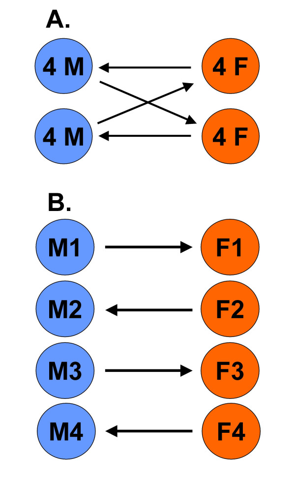
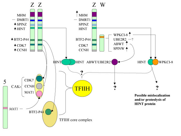
) indicate ≤2-fold difference, and long arrows (↑) indicate >2-fold difference, as determined by real-time PCR. Genes not differentially expressed are indicated with a dash (—). Long thin arrows (→) indicate gene product associations. A dotted bracket shows the possible interaction between HINT and the TFIIH complex.

References

    1. Morris JA, Jordan CL, Breedlove SM. Sexual differentiation of the vertebrate nervous system. Nat Neurosci. 2004;7:1034–1039. doi: 10.1038/nn1325. - DOI - PubMed
    1. Schantz SL, Widholm JJ. Cognitive effects of endocrine-disrupting chemicals in animals. Environ Health Perspect. 2001;109:1197–1206. - PMC - PubMed
    1. Arnold AP, Gorski RA. Gonadal steroid induction of structural sex differences in the central nervous system. Annu Rev Neurosci. 1984;7:413–442. doi: 10.1146/annurev.ne.07.030184.002213. - DOI - PubMed
    1. MacLusky NJ, Naftolin F. Sexual differentiation of the central nervous system. Science. 1981;211:1294–1302. - PubMed
    1. Dennis C. Brain development: the most important sexual organ. Nature. 2004;427:390–392. doi: 10.1038/427390a. - DOI - PubMed

Publication types

MeSH terms