. 2006;30(6):575-87.
doi: 10.1016/j.dci.2005.05.004. Epub 2005 Jun 20.

Evolution of antibody class switching: identification and transcriptional control of an Inu exon in the duck (Anas platyrhynchos)

Affiliations

Evolution of antibody class switching: identification and transcriptional control of an Inu exon in the duck (Anas platyrhynchos)

Mats L Lundqvist et al. Dev Comp Immunol. 2006.

Abstract

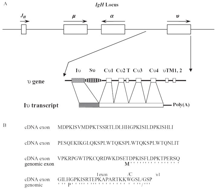
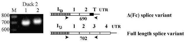
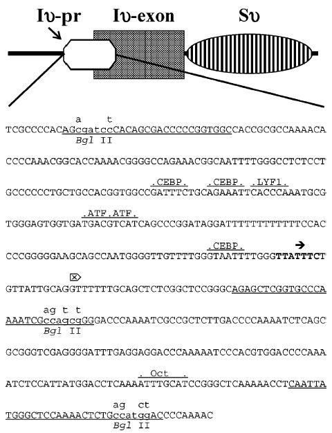
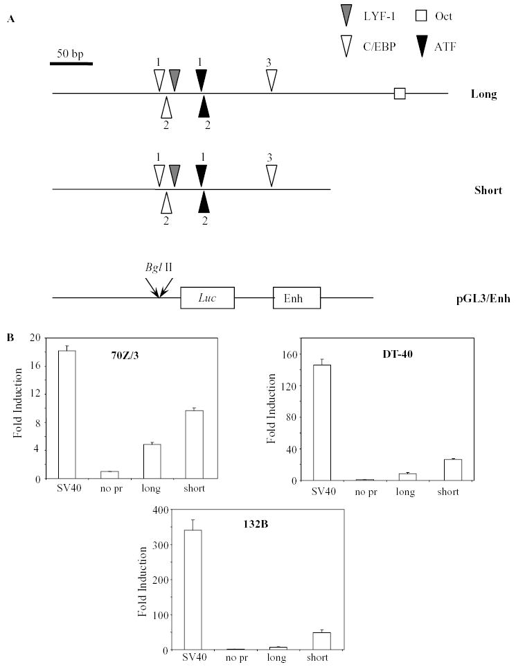
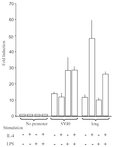
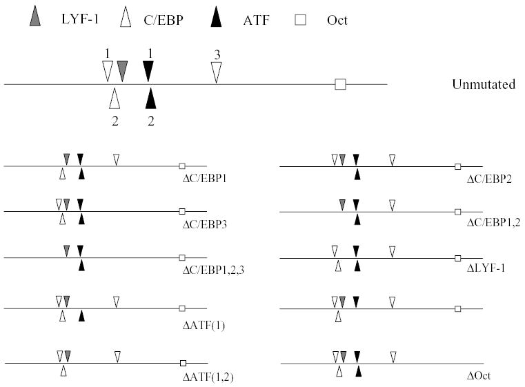
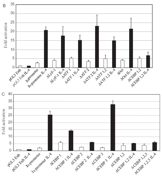
Immunoglobulin class switching is characteristic to the tetrapod lineage, but the nature of this process has been elucidated only in mammals, where I-exon transcription initiates and directs the recombination in the IgH locus. Here, it is shown that an I-exon occurs 5' of the nu (IgY constant region) gene of the duck (Anas platyrhynchos): it is longer than mammalian I-exons and comprised primarily of tandem repeats. The Inu promoter was identified and shown to be responsive to stimulation with IL-4 but not LPS. It contains Oct, LYF-1, ATF, and C/EBP motifs. Site directed mutagenesis indicates that 2 C/EBP motifs are uniquely necessary for the response of the promoter to IL-4, as tested in the mouse pre-B cell line, 70Z/3. These results support the conclusion that the signal transduction pathways controlling I-exon promoter responses to cytokines have been highly conserved in vertebrate evolution.


Figures

Fig. 1
Fig. 1
The Iυ exon of the duck: (A) Schematic showing the duck IgH locus and the relationship between the υ gene and the germ-line transcript, as cDNA. The position of the Iυ exon, the switch (Sυ) region, the constant (C) domain exons, the short terminal (T) exon and the transmembrane (TM) exons are indicated. The splicing event resulting in the characterized cDNA clone is shown. Figure is not drawn to scale. The corresponding positions in the duck genomic sequence (AJ314754) are for the I exon 41241–42218, and for the Cυ1 exon 45500–45784. The donor and acceptor sites for the Iυ–Cυ splice are TGG/GTATGG and CCACAG/GCA, respectively with the above mentioned positions in the genomic sequence; (B) Conceptual translation of the 3′ end of the Iυ exon examined as cDNA (this study) and from a genomic clone (21). The conceptual translations of the Iυ reading frame (defined by the Cυ1 to which the Iυ exon is spliced) were aligned from the 5′-most methionine codon (ATG) that excluded downstream stop codons. Amino acid identities are indicated as apostrophes (’). The differences in length and amino acid residues between the two sequences are inferred to reflect allelic polymorphism between the cDNA and genomic sequences examined. The Met codon for the genomic Iυ exon corresponds to position 42092–42094 in AJ314754.
Fig. 2
Fig. 2
Analysis (by agarose gel electrophoresis and ethidium bromide staining) of RT-PCR products of the Iυ germ-line transcripts (I/υ(ΔFc) and I/υ). Spleen mRNA from an immunized male duck (see Section 2) was used as template in the RT-PCR reaction. The amplicons were generated using a forward primer covering the Iυ/Cυ1 splice site, and reverse primers specific for the 3′ UT region of υ(ΔFc) (lane 2 and upper right image) and Cυ3 (lane 3 and lower right image). Lane 1 is a 100 bp ladder. The position of primers and the size of the predicted products (bp) are indicated on the schematic to the right of the gel, with the υ(ΔFc) form on top and the full length form below.
Fig. 3
Fig. 3
Schematic showing the relative positions of the Iυ promoter, Iυ exon and Sυ region (top). The sequence of the putative promoter region is shown below (pos. 40976–41470 in Acc no AJ314754). The identified transcription factor-binding sites are indicated above the sequence. Potential initiation regions (INRs, of consensus sequence YYANWYY, where N is the first nt transcribed) are shown in bold and arrow (→). PCR primers used to amplify the promoter region are underlined and lower case letters above the sequence indicates the BglII sites introduced to allow site specific cloning. The start of the cDNA clone (Acc. no. AJ517505) is shown with a (-[-graphic not found-]-).
Fig. 4
Fig. 4
Transcriptional activity of the Iυ promoter region: (A) Constructs of two different lengths (long and short), were generated by PCR and cloned in forward orientation into pGL3/Enh, a promoter-less luciferase reporter vector containing the SV40 enhancer; (B) The constructs were transiently transfected into the mouse pre-B cell line (70Z/3), and chicken B (DT40) and T (132B) cell lines. The pGL3 control vector, driven by the SV40 promoter and enhancer, was used as the positive control. The negative control was the pGL3/Enh vector. Each construct was transfected six times, and the luciferase activity of the constructs was normalized for transfection efficiency and expressed (mean ± standard deviation) as fold-induction compared to the negative control.
Fig. 5
Fig. 5
Induction of the Iυ promoter. The constructs (negative and positive controls, and the ‘long’ Iυ promoter in forward orientation) were transfected into the 70Z/3 mouse pre-B cell line. Post transfection, the cells were stimulated with LPS and IL-4 (indicated by + or − signs below the graph) either alone or in combination. Two experiments were conducted, in which each transfection/treatment was carried out three times. Both experiments showed the same effects, and the results of one experiment are shown. The activity of the constructs was normalized for transfection efficiency and expressed (mean ± SE) as fold-induction compared to the negative control.
Fig. 6
Fig. 6
C/EBP sites are necessary for the response of the Iυ promoter to IL-4: (A) Schematic representation of the native promoter and the sites mutated in each construct; (B) and (C) Transcriptional responses of the native and mutated Iυ promoter constructs. Transfections with no IL-4 stimulation (white bars) and with IL-4 stimulation (black bars) are shown as means ± standard deviation from one representative set of transfections in triplicate. The effects of the mutations on the response to IL-4 were calculated for three independent sets of triplicate transfections of each promoter construct and are displayed in Table 1.
Fig. 6
Fig. 6
C/EBP sites are necessary for the response of the Iυ promoter to IL-4: (A) Schematic representation of the native promoter and the sites mutated in each construct; (B) and (C) Transcriptional responses of the native and mutated Iυ promoter constructs. Transfections with no IL-4 stimulation (white bars) and with IL-4 stimulation (black bars) are shown as means ± standard deviation from one representative set of transfections in triplicate. The effects of the mutations on the response to IL-4 were calculated for three independent sets of triplicate transfections of each promoter construct and are displayed in Table 1.

Similar articles

References

    1. Moore KW, Rogers J, Hunkapiller T, Early P, Nottenburg C, Weissman I, et al. Expression of IgD may use both DNA rearrangement and RNA splicing mechanisms. Proc Natl Acad Sci USA. 1981;78(3):1800–4. - PMC - PubMed
    1. Stavnezer J, Kinoshito K, Muramatsu M, Honjo T. Molecular mechanism of class switch recombination. In: Honjo T, Alt FW, Neuberger M, editors. Molecular Biology of B Cells. London: Elsevier Academic Press; 2004. p. 307–26.
    1. Kinoshita K, Lee CG, Tashiro J, Muramatsu M, Chen XC, Yoshikawa K, et al. Molecular mechanism of immunoglobulin class switch recombination. Cold Spring Harb Symp Quant Biol. 1999;64:217–26. - PubMed
    1. Muramatsu M, Sankaranand VS, Anant S, Sugai M, Kinoshita K, Davidson NO, et al. Specific expression of activation-induced cytidine deaminase (AID), a novel member of the RNA-editing deaminase family in germinal center B cells. J Biol Chem. 1999;274(26):18,470–18,476. - PubMed
    1. Kemp DJ, Harris AW, Cory S, Adams JM. Expression of the immunoglobulin C mu gene in mouse T and B lymphoid and myeloid cell lines. Proc Natl Acad Sci USA. 1980;77(12):2876–80. - PMC - PubMed

Publication types

MeSH terms

Associated data