. 2006 May;17(5):2177-89.
doi: 10.1091/mbc.e05-10-0913. Epub 2006 Feb 15.

The Ras-GRF1 exchange factor coordinates activation of H-Ras and Rac1 to control neuronal morphology

Affiliations

The Ras-GRF1 exchange factor coordinates activation of H-Ras and Rac1 to control neuronal morphology

Huibin Yang et al. Mol Biol Cell. 2006 May.

Abstract

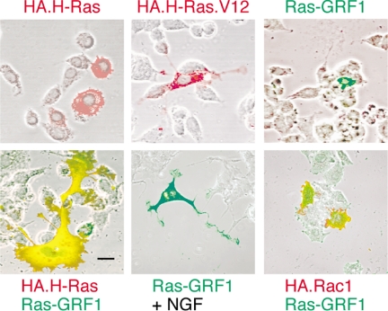
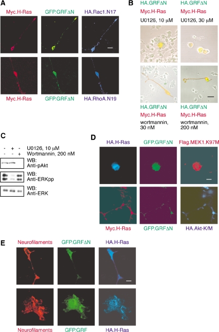
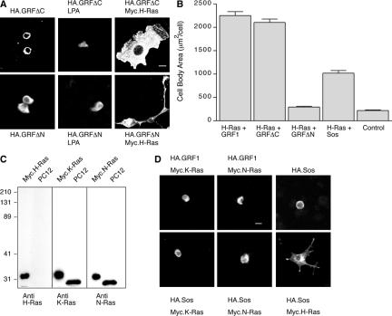
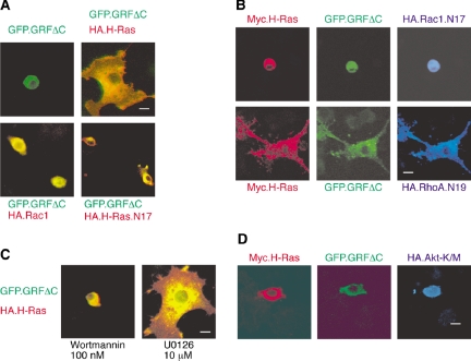
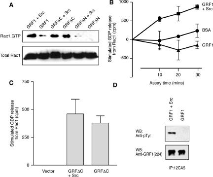
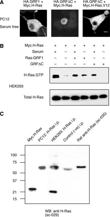
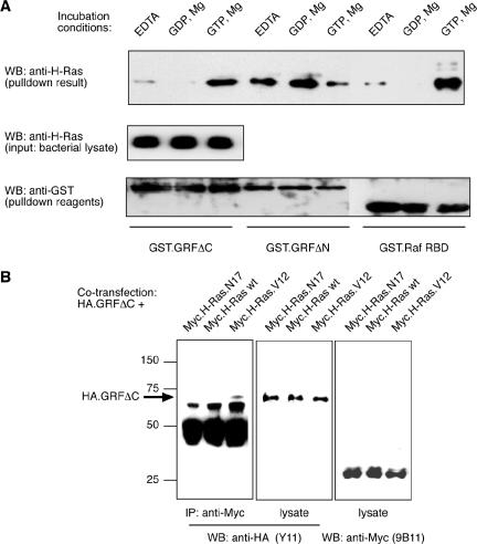
The Ras-GRF1 exchange factor has regulated guanine nucleotide exchange factor (GEF) activity for H-Ras and Rac1 through separate domains. Both H-Ras and Rac1 activation have been linked to synaptic plasticity and thus could contribute to the function of Ras-GRF1 in neuronal signal transduction pathways that underlie learning and memory. We defined the effects of Ras-GRF1 and truncation mutants that include only one of its GEF activities on the morphology of PC12 phaeochromocytoma cells. Ras-GRF1 required coexpression of H-Ras to induce morphological effects. Ras-GRF1 plus H-Ras induced a novel, expanded morphology in PC12 cells, which was characterized by a 10-fold increase in soma size and by neurite extension. A truncation mutant of Ras-GRF1 that included the Ras GEF domain, GRFdeltaN, plus H-Ras produced neurite extensions, but did not expand the soma. This neurite extension was blocked by inhibition of MAP kinase activation, but was independent of dominant-negative Rac1 or RhoA. A truncation mutant of Ras-GRF1 that included the Rac GEF domains, GRFdeltaC, produced the expanded phenotype in cotransfections with H-Ras. Cell expansion was inhibited by wortmannin or dominant-negative forms of Rac1 or Akt. GRFdeltaC binds H-Ras.GTP in both pulldown assays from bacterial lysates and by coimmunoprecipitation from HEK293 cells. These results suggest that coordinated activation of H-Ras and Rac1 by Ras-GRF1 may be a significant controller of neuronal cell size.


Figures

Figure 1.
Figure 1.
Effects of Ras-GRF1 expression on the morphology of PC12 cells. PC12 cells were transfected to express the indicated constructs for 48 h. Expression of Ras-GRF1 was detected by green indirect polyclonal immunofluorescence. Expression of HA-tagged H-Ras, H-Ras.V12, or Rac1 were detected by red indirect monoclonal immunofluorescence for the HA tag. To compare morphology with untransfected cells, the fluorescence results are overlaid with a phase-contrast image of the field. Where indicated, the cultures were treated with 40 ng/ml NGF (Genentech) to induce neurite extension (bottom middle). Scale bar, 50 μm. Pictures shown are typical of results from a minimum of three independent experiments. The proportion of cells cotransfected with both H-Ras and Ras-GRF1 (bottom left) that exhibited the novel, expanded morphology was ∼25%. This morphology was never detected after transfection with either H-Ras or Ras-GRF1 alone.
Figure 2.
Figure 2.
GRFΔN/H-Ras–induced neurite extension is dependent on ERK activation but not PI3-Kinase or Rho proteins. (A) Triple transfections were performed in PC12 cells, using GFP.GRFΔN (green fluorescent detection), Myc.H-Ras (red indirect immunofluorescent detection), and a dominant-negative variant of either Rac1 (HA.Rac1.N17) or RhoA (HA.RhoA.N19; blue indirect immunofluorescent detection). The result shown is of two single, triply transfected cells that are representative of multiple cells from three independent experiments. Scale bar, 50 μm. Neurite outgrowth of PC12 cells was unaffected by the presence of either dominant-negative Rac1 (top row) or RhoA (bottom row). (B) PC12 cells transfected with HA.GRFΔN (green detection) and Myc.H-Ras (red detection) were maintained in culture medium in the presence of U0126, a MEK inhibitor, or wortmannin, a PI3K inhibitor, for 48 h. U0126 at 10 μM was sufficient to inhibit neurite extension of PC12 cells (top panels). In contrast, neurite outgrowth was not blocked by wortmannin (bottom panels). The results are representative of three independent experiments. (C) Lysates were prepared from PC12 cells that had been treated for 1 h with DMSO vehicle, or 10 μM U0126, or 200 nM wortmannin and subjected to Western blotting for phosphorylated Akt, dually phosphorylated ERK MAP kinases and total ERK as previously described (Mattingly et al., 2001b; Menard et al., 2005). (D) Triple transfections were performed using GFP.GRFΔN (green fluorescent detection), HA- or Myc-tagged H-Ras (blue and red indirect immunofluorescent detection), and dominant-negative variants of either MEK1 (Flag.MEK1.K97M) or Akt (HA.Akt-K/M) (red and blue indirect immunofluorescent detection). The result shown is of two representative, triply transfected cells. Scale bar, 50 μm. Neurite outgrowth from PC12 cells was completely blocked by dominant-negative MEK1 (top row) but unaffected by dominant-negative Akt (bottom row). (E) Cotransfections were performed in PC12 cells, using GFP.GRFΔN or GFP.GRF1 (green fluorescent detection), and HA.H-Ras (blue indirect immunofluorescent detection). PC12 cells that differentiate in response to coexpression of GFP.GRFΔN plus H-Ras (top row) or GFP.Ras-GRF1 plus H-Ras (bottom row) are positive for expression of neurofilaments (red indirect immunofluorescent detection).
Figure 3.
Figure 3.
The expanded PC12 morphology can be induced by cotransfection of GRFΔC and H-Ras. (A) Transfection with either HA.GRFΔC (left, top panel) or HA.GRFΔN (left, bottom panel) alone did not alter cell body size of PC12 cells. Treatment of the cells with 10 μM LPA (middle panels) did not stimulate any morphological differentiation. The combination of HA.GRF1ΔC and Myc.H-Ras produced the expanded morphology (right, top panel). Scale bar, 50 μm. The combination of HA.GRFΔN and Myc.H-Ras produced cells with small soma but long neurite extensions (right, bottom panel). (B) Using the Metamorph software program, we measured cell body areas of transfected cells. We measured 75 cells within each group that were positive for both transfected proteins or else negative for both (as control) from three independent experiments. The largest 10 measurements for each condition from each experiment (total of 30 cells) were averaged to determine cell body area (mean ± SEM). The ability of Sos1 plus H-Ras to increase cell soma size was significantly less than that of Ras-GRF1 plus H-Ras (p < 0.001, by two-way analysis of variance). (C) PC12 cell lysate was separated on 12% SDS-polyacrylamide gel in parallel with positive controls of Myc-tagged Ras proteins expressed in COS-7 cells and transferred onto nitrocellulose membrane. Immunoblotting of the standards with anti-Myc reagents verified their relative loading (Mattingly et al., 2006). The blots were then probed with anti-H-Ras, anti-K-Ras, or anti-N-Ras monoclonal antibodies, as shown. The result showed that the PC12 cells express K-Ras and N-Ras but no detectable H-Ras. (D) Transfection of PC12 cells with Ras-GRF1 plus K-Ras or N-Ras, or with Sos1 alone (top row), or with Sos1 plus K-Ras or N-Ras (bottom left and middle) did not induce morphological changes. Transfection of PC12 cells with Sos1 and H-Ras produced some neurite extension and expansion of the soma (bottom right).
Figure 4.
Figure 4.
The expanded PC12 morphology that is induced by GRFΔC/H-Ras is blocked by dominant-negative Rac1 as well as inhibition of PI 3-kinase. (A) PC12 cells were transfected with either GFP.GRFΔC (green fluorescent detection) alone or in combination with HA-tagged H-Ras, H-Ras.N17, or Rac1 (red indirect immunofluorescent detection). The expanded morphology induced by GFP.GRFΔC requires H-Ras (right, top panel). Scale bar, 50 μm. GFP.GRFΔC alone as well as its combination either with a dominant-negative H-Ras variant, H-Ras.N17, or with Rac1 did not induce changed morphology. (B) Triple transfection of GFP.GRFΔC (green detection) and Myc-H-Ras (red detection) plus either HA-Rac1.N17 or HA-RhoA.N19 (blue indirect immunofluorescent detection) was performed in PC12 cells. The expanded morphology induced by GRFΔC/H-Ras, was blocked by Rac1.N17 (top row), but not by RhoA.N19 (bottom row). (C) GFP.GRFΔC and H-Ras exhibit a great expansion of the soma. Expansion of the soma is effectively blocked by wortmannin, a PI3K inhibitor (left panel), but not by U0126, a MEK inhibitor (right panel). (D) Coexpression of dominant-negative HA-Akt-K/M (blue indirect immunofluorescent detection) with GFP.GRFΔC (green detection) and Myc-H-Ras (red detection) blocked expansion of the soma.
Figure 5.
Figure 5.
Activation of Rac1 by GRF1/Src correlates with tyrosine phosphorylation of GRF1. (A) HEK-293 cells were transfected with HA-tagged GRF1, GRFΔC, or GRFΔN, with or without additional activated Src. Activation of the endogenous Rac1 protein was then assayed by determination of the proportion that was bound to GTP. (B) HEK-293 cells were transfected with HA.GRF1 alone or in combination with activated Src. Cell lysates were prepared after serum starvation and anti-HA immunoprecipitates were assayed for their ability to release [3H]GDP that had been prebound to recombinant Rac1 in comparison to incubations with BSA as a control. Data shown are mean ± SEM from four independent experiments conducted in triplicate. (C) HEK-293 cells were transfected with a control, empty vector, or with HA.GRF1ΔC alone or plus activated Src. Anti-HA immunoprecipitates were assayed for 20 min for their ability to release [3H]GDP that had been prebound to recombinant Rac1. Data shown are the change in released GDP relative to that from Rac1 incubated with a control immunoprecipitate and are mean ± SEM from four independent experiments conducted in triplicate. (D) HEK-293 cells transfected with either HA.GRF1 alone or in combination with Src. After serum starvation, cell lysates were prepared in RIPA buffer as described (Yang et al., 2003). HA.Ras-GRF1 was immunoprecipitated and analyzed by Western blot with anti-pTyr antibody (top panel). The blot was then stripped and reprobed with anti-GRF1 antibody sc224 (bottom panel).
Figure 6.
Figure 6.
Ras-GRF1 can activate H-Ras and induce expanded PC12 morphology independent of serum; GRF1ΔC requires activation of H-Ras. (A) PC12 cells were transfected with HA.GRF1 in combination with Myc.H-Ras, or with HA.GRF1ΔC in combination either with Myc.H-Ras or Myc.H-Ras.V12 and then cultured for 48 h in the absence of serum. Cells were fixed and subjected to indirect immunofluorescence staining using anti-HA polyclonal antibody Y11 and green detection or anti-Myc mAb 9e10 and red detection. In the absence of serum, GRF1 and H-Ras could still induce cell expansion (left) but GRF1ΔC plus H-Ras could not (center). A combination of GRF1ΔC plus H-Ras.V12 did induce cell expansion (right). Scale bar, 50 μm. (B) HEK-293 cells were transfected with Myc.H-Ras alone, or in combination with Ras-GRF1 or with GRF1ΔC. Ras.GTP pulldown assays were performed on the cell lysates. The active form of the transfected H-Ras was detected by Western blot using anti-Myc 9B11 antibody (top panel). Cell lysates were probed for total transfected H-Ras to determine whether there were equivalent amounts of Myc.H-Ras protein in each sample (bottom panel). (C) Lysates of PC12 and HEK293 cells were immunoprecipitated for H-Ras using a rat anti-H-Ras mAb, followed by rabbit anti-rat polyclonal antibodies and protein A-Sepharose. The immunoprecipitates were analyzed by Western blot in comparison to a Myc-tagged H-Ras positive control, and negative controls of an immunoprecipitation from HEK293 cell lysate that omitted the rat anti-H-Ras antibody and of the rat anti-H-Ras antibody alone.
Figure 7.
Figure 7.
Ras-GRF1ΔC binds preferentially to GTP-bound H-Ras in a GST-fusion protein pulldown assay and in a coimmunoprecipitation. (A) Lysates of bacteria expressing H-Ras bacteria were pretreated with 5 mM EDTA, 5 mM magnesium plus 100 μM GTP, or GDP, and then incubated with glutathione beads coated with GST.GRFΔC, GST.GRFΔN, or GST.RafRBD. The results shown are for the H-Ras that was detected by Western blotting of the washed pulldown reactions (top panel) or of 5% of the bacterial lysates (middle panel). The membranes were stripped and reprobed with anti-GST antibody to control for the relative loading of the GST fusion proteins onto the pulldown beads (bottom panel). Results shown are representative of three independent experiments. (B) HEK-293 cells were cotransfected with HA.GRFΔC plus Myc.H-Ras, Myc.H-Ras.N17, or Myc.H-Ras.V12. Cell lysates were prepared and blotted for content of Myc-tagged H-Ras proteins (right panel) and HA.GRFΔC (center panel) that confirmed even expression of the constructs. Anti-Myc immunoprecipitates were prepared from cell lysates using the mAb 9B11. HA.GRFΔC was only detected in immunoprecipitations from cells that coexpressed Myc.H-Ras.V12 (left panel).
Figure 8.
Figure 8.
The expanded PC12 cell morphology that is induced by Ras-GRF1 is produced by coordinated activation of H-Ras and Rac1. Expansion of the PC12 soma requires activation of both H-Ras and Rac1. Full-length Ras-GRF1 can accomplish both functions, but GRFΔC requires an additional stimulus to activate H-Ras. Activated H-Ras can directly interact with GRFΔC and may provide a mechanistic link for coordination of Rac1 activation at the locus of H-Ras activation.

Similar articles

Cited by

References

    1. Arendt, T. et al. (2004). Neuronal activation of Ras regulates synaptic connectivity. Eur. J. Neurosci. 19, 2953–2966. - PubMed
    1. Arozarena, I., Matallanas, D., Berciano, M. T., Sanz-Moreno, V., Calvo, F., Munoz, M. T., Egea, G., Lafarga, M., and Crespo, P. (2004). Activation of H-Ras in the endoplasmic reticulum by the RasGRF family guanine nucleotide exchange factors. Mol. Cell. Biol. 24, 1516–1530. - PMC - PubMed
    1. Aznar, S., and Lacal, J. C. (2001). Rho signals to cell growth and apoptosis. Cancer Lett. 165, 1–10. - PubMed
    1. Baouz, S., Jacquet, E., Bernardi, A., and Parmeggiani, A. (1997). The N-terminal moiety of CDC25(Mm), a GDP/GTP exchange factor of Ras proteins, controls the activity of the catalytic domain. Modulation by calmodulin and calpain. J. Biol. Chem. 272, 6671–6676. - PubMed
    1. Bar-Sagi, D., and Feramisco, J. R. (1985). Microinjection of the Ras oncogene protein into PC12 cells induces morphological differentiation. Cell 42, 841–848. - PubMed

Publication types

MeSH terms

Substances