. 2006 Feb 15;26(7):1896-905.
doi: 10.1523/JNEUROSCI.3202-05.2006.

Genetic analyses demonstrate that bone morphogenetic protein signaling is required for embryonic cerebellar development

Affiliations

Genetic analyses demonstrate that bone morphogenetic protein signaling is required for embryonic cerebellar development

Lihua Qin et al. J Neurosci. .

Abstract

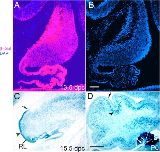
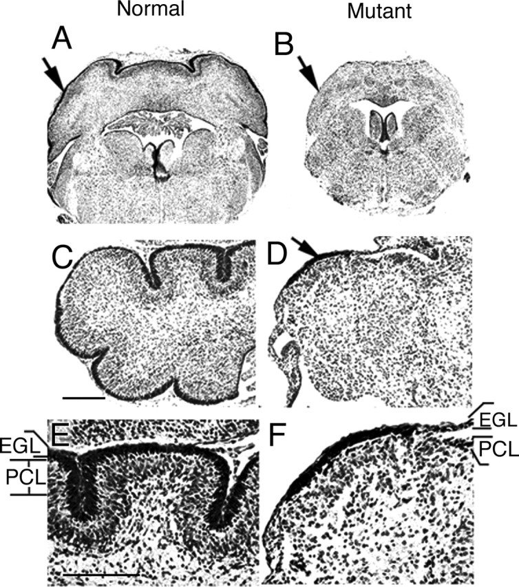
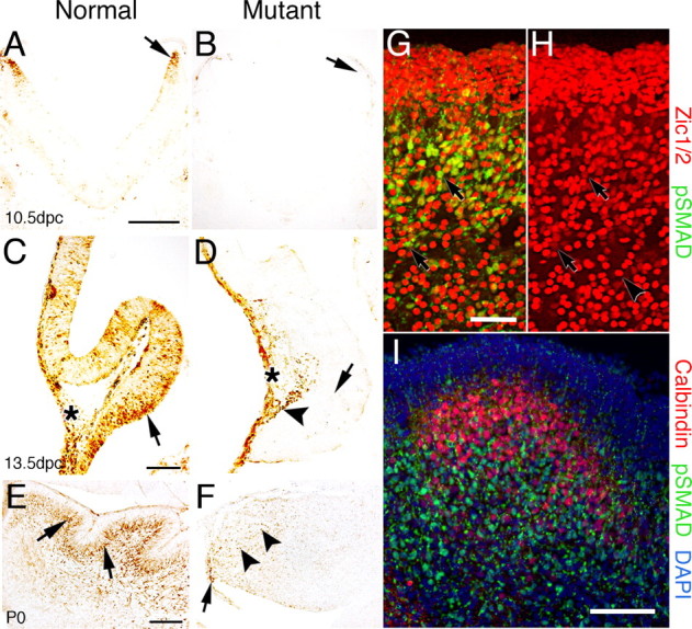
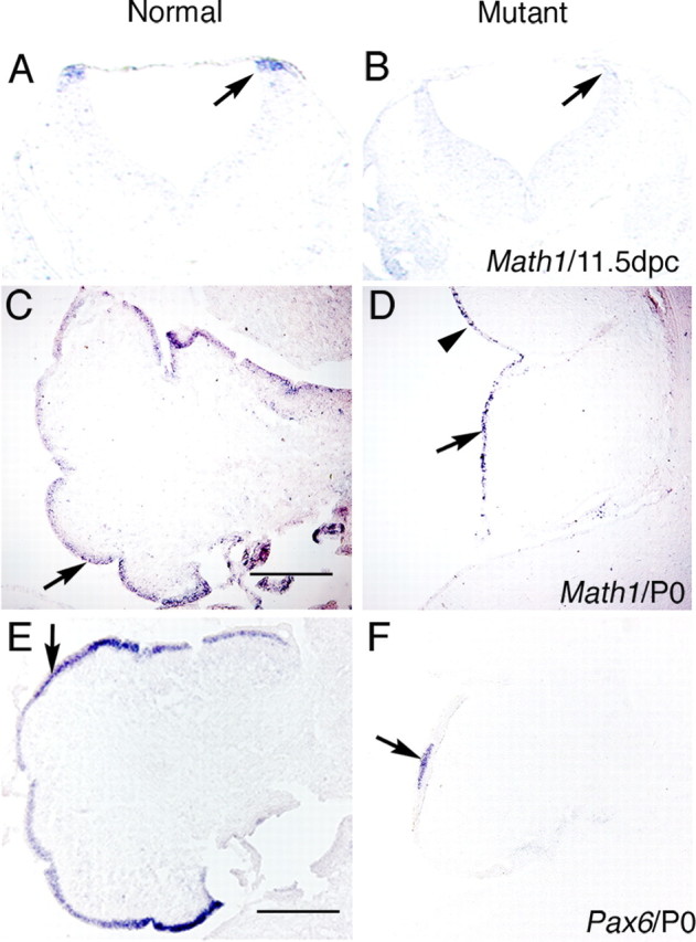
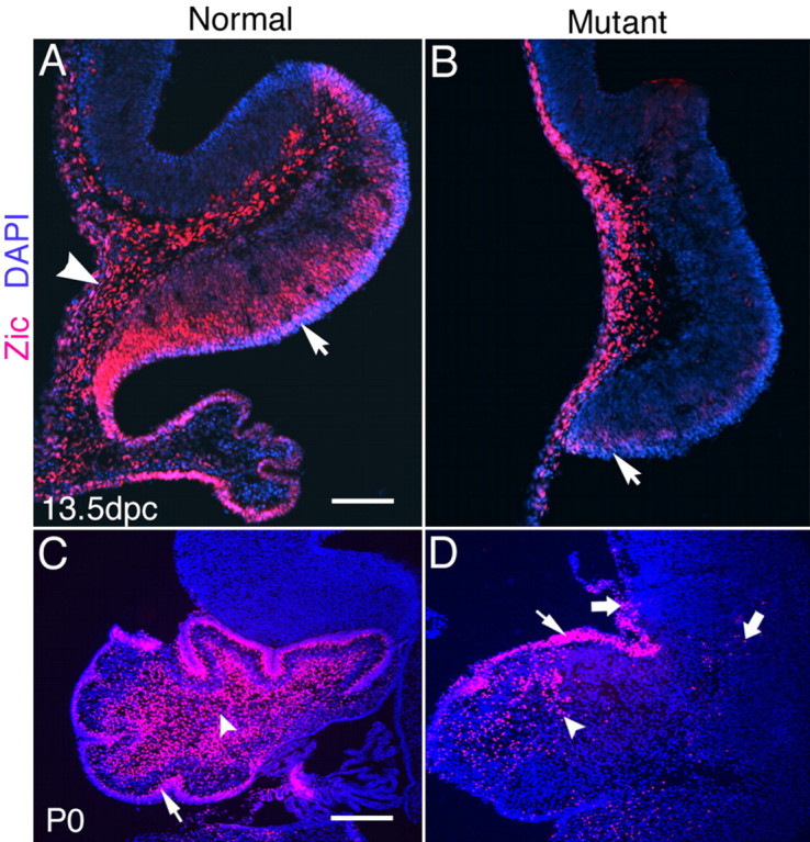
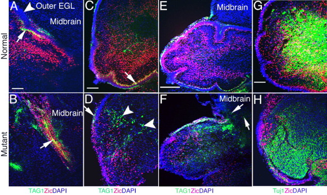
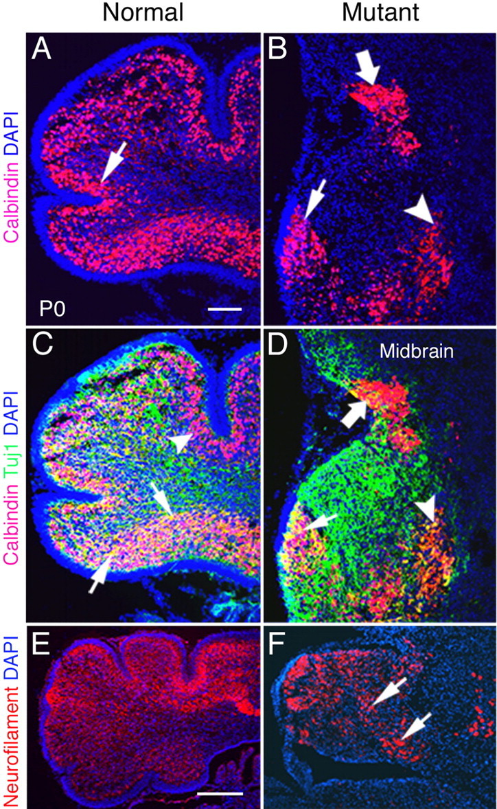
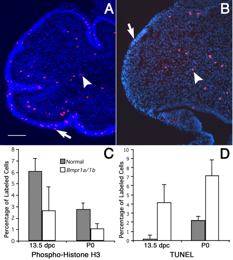
The cerebellum has been a useful model for studying many aspects of neural development because of its relatively simple cytoarchitecture and developmental program. Yet, the genetic mechanisms underlying early differentiation and patterning of the cerebellum are still poorly characterized. Cell expression studies and culture experiments have suggested the importance of bone morphogenetic proteins (BMPs) in development of specific populations of cerebellar neurons. Here, we examined mice with targeted mutations in the BMP type I receptor genes Bmpr1a and Bmpr1b, to genetically test the hypothesis that BMPs play an inductive role in the embryogenesis of cerebellar granule cells. In Bmpr1a;Bmpr1b double knock-out mice, severe cerebellar patterning defects are observed resulting in smaller cerebella that are devoid of foliation. In mutants containing either single BMP receptor gene mutation alone, cerebellar histogenesis appears normal, thereby demonstrating functional redundancy of type I BMP receptors during cerebellar development. Loss of BMP signaling in double mutant animals leads to a dramatic reduction in the number of cerebellar granule cells and ectopic location of many of those that remain. Molecular markers of granule cell specification, including Math1 and Zic1, are drastically downregulated. In addition, Purkinje cells are disorganized and ectopically located, but they appear to be correctly specified. Consistent with the interpretation that granule cells alone are affected, phosphorylated Smad1/5/8 is immunolocalized predominantly to granule cell precursors and not appreciably detected in Purkinje cell precursors. This study demonstrates that BMP signaling plays a crucial role in the specification of granule cells during cerebellar development.


Figures

Figure 1.
Figure 1.
Cre-mediated recombination of the ROSA26 reporter by the Bcre-32 transgene is detected in the cerebellum. A, At 13.5 dpc, immunohistochemical staining for β-galactosidase (β-Gal) shows lacZ expression in the vast majority of cells throughout the cerebellar anlage. B, DAPI staining shows the density of cells within the cerebellum. C, At 15.5 dpc, Cre-mediated lacZ expression is detected strongly in the EGL, because of the high cell density in this region (arrowhead). Note that the EGL extends from the rhombic lip (RL) but only covers part of the cerebellar cortex at this stage of development (ending at the arrow). D, Postnatally, Cre-mediated lacZ expression is found in the EGL (arrow) and the PCL (arrowhead). Scale bars: B, 100 μm; D, 250 μm.
Figure 2.
Figure 2.
Abnormal formation/development of the cerebellum in Bmpr1a;Bmpr1b double knock-out mutants. A, Nissl staining on coronal sections shows the normal cerebellum at P0 (arrow). B, The cerebellum of the mutant mouse is smaller in size at P0 (arrow). C, E, Nissl staining show defined layers including the EGL and PCL in normal P0 neonates. The ML is the cell-sparse layer between the EGL and PCL. D, F, In the mutant cerebellum, disruption of the layered organization is observed. Only a small region with the thickened EGL (arrow) is observed, but most of the mutant cerebellum completely lacks the EGL and PCL, the presumptive position of which is indicated in F. E, High-magnification view of C. F, High-magnification view of D. Scale bars: C, E, 250 μm.
Figure 3.
Figure 3.
Phospho-SMAD immunolabeling demonstrates loss of BMP signaling in the cerebellum of Bmpr1a;Bmpr1b knock-out mice. A, Immunoreactive cells are detected in the rostral rhombic lip at 10.5 dpc in normal mice (arrow). B, In mutant embryos, phospho-SMAD immunostaining is lost in the rostral rhombic lip (arrow). C, At 13.5 dpc in normal embryos, immunoreactive cells are detected in the ventral cerebellar anlage within and above the neuroepithelium (arrow). In addition, immunoreactive cells are located dorsally in leptomeninges (asterisk). D, In the mutant mouse, phospho-Smad immunostaining demonstrates that BMP signaling has been abrogated in the vast majority of cells within the cerebellar anlage (arrow). However, a few positive cells are located along the dorsal side (arrowhead) of the neural tube in a region where the granule cells are migrating to form the EGL. Immunolabeling in the leptomeninges is unaffected (asterisk). E, Postnatally, phospho-SMAD-immunolabeled cells are detected in migrating granule cells and in the IGL of normal mice, which lies deep to the PCL (arrows). F, In the P0 mutant, phospho-Smad immunolabeling is drastically reduced. However, a few labeled cells are detected within or proximal to the rhombic lip (arrow) and a few cells deep in the cerebellum (arrowheads). G, Double immunolabeling with phospho-SMAD (green) and Zic1/2 (red) in the cerebellum of a normal mouse at P0. Double immunostained cells are located predominantly in the IGL and in migrating granule cells (arrows). However, a few cells are detected in the EGL (the dense cell layer located at the top of the panel). H, Area depicted in G showing immunostaining for Zic1/2 alone to demonstrate that two classes of Zic1/2-immunopositive cells can be detected that label intensely (arrowhead) or lightly (arrows). I, Double immunolabeling with phospho-SMAD (green) and calbindin (red) in a section of the cerebellum of normal mice at P0 counterstained with DAPI (blue). Virtually no double immunostained cells are detected in the cerebella, whereas single calbindin-labeled positive Purkinje cells are found in the cerebella. These results demonstrate that phospho-SMAD is detected in granule cells that have migrated away from the proliferative zone of the EGL and are in the process of differentiating into mature granule cells. Scale bars: A, 250 μm; C, E, I, 100 μm; G, 50 μm. pSMAD, Phospho-SMAD.
Figure 4.
Figure 4.
Expression of specific markers demonstrates granule cell loss in BMP signaling mutants at 11.5 dpc and P0. A–D, In situ hybridization showing Math1 expression demonstrates loss of granule cell precursors in the rostral rhombic lip and cerebellum of Bmpr1a;Bmpr1b double knock-out mutants. A, Math1 expression is found in the rostral rhombic lip at 11.5 dpc in normal embryos (arrow). B, Math1 expression is lost at 11.5 dpc in mutant animals (arrow). C, Math1 expression is detected in the EGL at P0 in normal embryos (arrow). D, At P0, Math1 expression is detected in a reduced number of cells that are distributed along the entire rim of the cerebellar cortex where the EGL would normally form (arrow) and into the midbrain, which does not normally express Math1 (arrowhead). E, Pax6 expression is detected in the EGL of normal embryos at P0 (arrow). F, In mutants at P0, Pax6 expression is lost in most regions with the exception of a small region that corresponds to the region of the thickened, more normal EGL observed in the mutants (arrow). Scale bars: C, E, 250 μm.
Figure 5.
Figure 5.
Expression of Zic1/2 in the cerebellum at 13.5 dpc and P0. A, Zic1/2-labeled cells form a wide layer in the ventral side of the cerebellum in the normal mouse at 13.5dpc (arrow). Expression is also detected in the leptomeninges covering the neural tube and the choroid plexus (arrowhead). B, In 13.5 dpc mutant embryos, Zic1/2-labeled cells are detected predominantly in the leptomeninges and extra-neural tube tissues, except for light staining just above the neuroepithelium on the ventral side of the cerebellum (arrow). C, Zic1/2-labeled cells are found in the inner EGL (arrow) and deep in the cerebellum in normal mice at P0 (arrowhead). The vast majority of Zic1/2-labeled cells are localized in the cerebellum of normal embryos. D, The number of Zic1/2-labeled cells is drastically decreased both in the EGL (small arrow) and deep in the cerebellum (arrowhead) of the double knock-outs at P0. Some Zic1/2-labeled cells migrate into the mesencephalon in mutants (large arrows). Scale bars: A, 100 μm; C, 250 μm.
Figure 6.
Figure 6.
Differentiation of postmitotic granule cells is disturbed in double knock-outs. A–F, Double immunostaining with antibodies directed against TAG-1 (green) and Zic1/2 (red) at P0 in normal and mutant neonates. A, TAG-1-labeled fibers are detected in the inner EGL and form a layer in the superior edge in normal neonates (arrow). The outer EGL has no labeled fibers (arrowhead). B, TAG-1-labeled fibers mix with Zic1/2-positive cells in the superior edge in the double knock-out mice (arrow). C, TAG-1-labeled fibers are also found in the inner EGL at the ventral side of the normal cerebellum (arrow). D, TAG-1-labeled fibers are detected in the deep cerebellum in clusters (arrowheads) with only a small region appearing more normal in mutant cerebellum (arrow). E, TAG-1-labeled fibers are restricted to the cerebellum in the normal mouse and are not detected in the midbrain. F, TAG-1-labeled fibers (arrows) and Zic1/2-positive cells are found in the midbrain of the mutant. G, H, Double immunolabeling for Tuj1 (green) and Zic1/2 (red) in the cerebellum at P0. G, Tuj1 expression and more double-labeled cells are located in the deep layers of the normal cerebellum. H, Decreased Tuj1 expression and few double-labeled cells are found in the mutant cerebellum. Scale bars: A, C, G, 100 μm; E, 250 μm.
Figure 7.
Figure 7.
Maturation and migration of Purkinje cells are abnormal in P0 Bmpr1a;Bmpr1b knock-outs. A, At P0, most calbindin-positive Purkinje cells are localized beneath the EGL and form a distinct layer in normal animals (arrow). B, However, in mutants, most of the Purkinje cells are localized ectopically in the deep cerebellum (arrowhead) and midbrain (large arrow). Only a small number of cells are localized correctly (arrow). C, D, Detection of calbindin (red) and Tuj1 (green) immunolabeling at P0. C, Double-labeled Purkinje cells are detected in the ventral side of the cerebellum in normal neonates (arrows). Purkinje cells labeled with calbindin alone are detected in the dorsal side of the cerebellum (arrowhead). D, Double-labeled cells are located ectopically in the deep cerebellum (arrowhead) and are present in decreased number in the mutant neonates. Tuj1 expression is disorganized, and some double-labeled cells are located in the midbrain of double knock-out neonates (large arrow). Only some double-labeled cells are localized correctly (small arrow). E, In normal P0 mice, the majority of cerebellar cells express neurofilament, except those located in the EGL. F, Neurofilament expression is detected in the cerebellum in plaques (arrows) with some areas lacking expression in double mutants. Scale bars: A, 100 μm; E, 250 μm.
Figure 8.
Figure 8.
Decreased proliferation and increased apoptosis in Bmpr1a;Bmpr1b double knock-out mutants. A, B, Phospho-histone H3 immunolabeling detects proliferating cells at P0. A, Phospho-histone H3-labeled cells are found in the EGL (arrow) and deep cerebellum (arrowhead) in normal mice. Scale bar, 100 μm. B, In double knock-out mutants, the number of phospho-histone H3-positive cells decreases markedly, both in the EGL (arrow) and the deep cerebellum (arrowhead). C, The number of proliferating cerebellar cells decreases by 2.3-fold (p < 0.004) at 13.5 dpc and by 2.6-fold (p < 0.0001) in the deep cerebellum at P0. D, A significant 27-fold increase in cell death is observed in the mutant (p < 0.0003) at 13.5 dpc, whereas the TUNEL-positive cells increase 3.3-fold in the mutant at P0 (p < 0.0001). Statistical tests of significance were accomplished using a one-tailed Student’s t test. Error bars indicate SD.

Similar articles

Cited by

References

    1. Ahn K, Mishina Y, Hanks MC, Behringer RR, Crenshaw EB III (2001). BMPR-IA signaling is required for the formation of the apical ectodermal ridge and dorsal-ventral patterning of the limb. Development 128:4449–4461. - PubMed
    1. Akazawa C, Ishibashi M, Shimizu C, Nakanishi S, Kageyama R (1995). A mammalian helix-loop-helix factor structurally related to the product of Drosophila proneural gene atonal is a positive transcriptional regulator expressed in the developing nervous system. J Biol Chem 270:8730–8738. - PubMed
    1. Alder J, Cho NK, Hatten ME (1996). Embryonic precursor cells from the rhombic lip are specified to a cerebellar granule neuron identity. Neuron 17:389–399. - PubMed
    1. Alder J, Lee KJ, Jessell TM, Hatten ME (1999). Generation of cerebellar granule neurons in vivo by transplantation of BMP-treated neural progenitor cells. Nat Neurosci 2:535–540. - PubMed
    1. Amy WH, Katherine G, Andrew A, Trisha S, Jane EJ (2001). Overexpression of Math1 disrupts the coordination of neural differentiation in cerebellum development. Mol Cell Neurosci 17:671–682. - PubMed

MeSH terms

Substances

LinkOut - more resources