. 2006 Feb 15;26(7):1923-34.
doi: 10.1523/JNEUROSCI.4359-05.2006.

Matrix metalloproteinase-9 is required for hippocampal late-phase long-term potentiation and memory

Affiliations

Matrix metalloproteinase-9 is required for hippocampal late-phase long-term potentiation and memory

Vanja Nagy et al. J Neurosci. .

Abstract

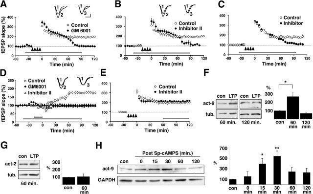
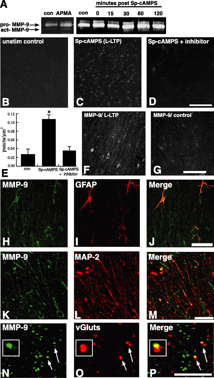
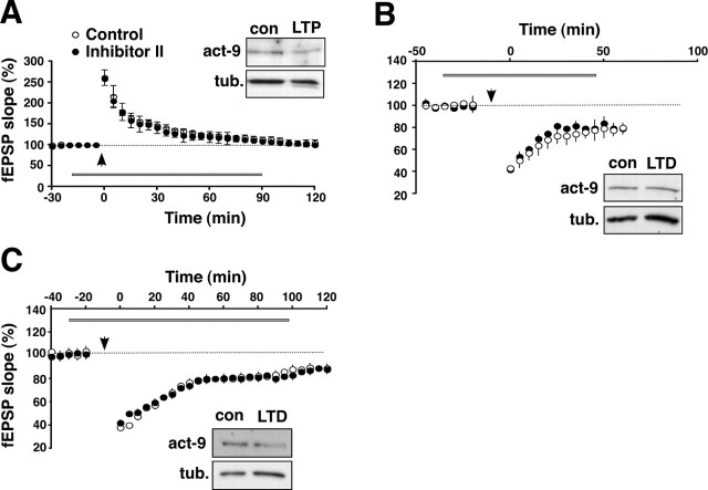
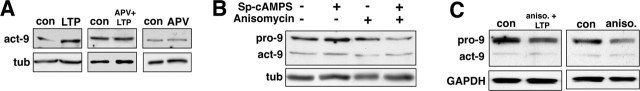
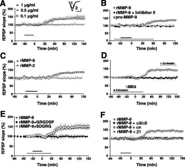
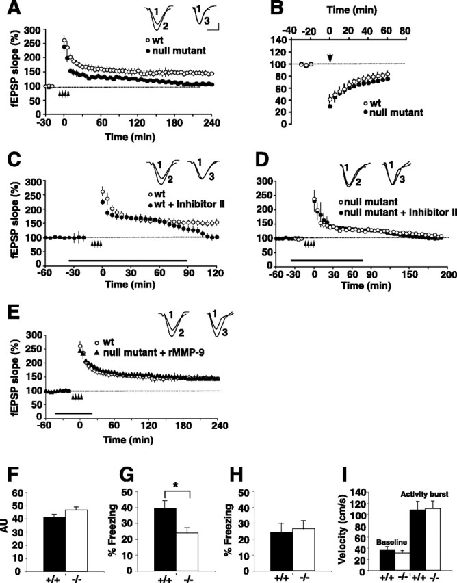
Matrix metalloproteinases (MMPs) are extracellular proteases that have well recognized roles in cell signaling and remodeling in many tissues. In the brain, their activation and function are customarily associated with injury or pathology. Here, we demonstrate a novel role for MMP-9 in hippocampal synaptic physiology, plasticity, and memory. MMP-9 protein levels and proteolytic activity are rapidly increased by stimuli that induce late-phase long-term potentiation (L-LTP) in area CA1. Such regulation requires NMDA receptors and protein synthesis. Blockade of MMP-9 pharmacologically prevents induction of L-LTP selectively; MMP-9 plays no role in, nor is regulated during, other forms of short-term synaptic potentiation or long-lasting synaptic depression. Similarly, in slices from MMP-9 null-mutant mice, hippocampal LTP, but not long-term depression, is impaired in magnitude and duration; adding recombinant active MMP-9 to null-mutant slices restores the magnitude and duration of LTP to wild-type levels. Activated MMP-9 localizes in part to synapses and modulates hippocampal synaptic physiology through integrin receptors, because integrin function-blocking reagents prevent an MMP-9-mediated potentiation of synaptic signal strength. The fundamental importance of MMP-9 function in modulating hippocampal synaptic physiology and plasticity is underscored by behavioral impairments in hippocampal-dependent memory displayed by MMP-9 null-mutant mice. Together, these data reveal new functions for MMPs in synaptic and behavioral plasticity.


Figures

None
MMP-9 is required for, and upregulated during, L-LTP in area CA1. A, Young rat hippocampal slices were subjected to an L-LTP-inducing tetanic stimulation protocol (arrows; four trains of 100 Hz, 1 s stimulation separated by 5 min), which produces both an E-LTP and a long-lasting L-LTP (open circles). In contrast, in the presence of GM6001 (25 μm), a broad-spectrum MMP inhibitor (bar), normal E-LTP is elicited but L-LTP is abolished, as shown by the return of the trace to baseline levels between 60 and 90 min (filled circles; n = 6 rats). Inset, Representative EPSP traces were recorded before tetanus (1), 90 min after tetanus (2), and 90 min after tetanus in the presence of GM6001 (3). B, Young rat hippocampal slices exposed to an MMP-2/9 inhibitor (Inhibitor II; 50 μm) and subjected to L-LTP-inducing tetanic stimulation also show normal E-LTP but abolishment of L-LTP (filled circles). Other conventions are as in A. Inset, Representative EPSP traces were recorded before tetanus (1), 90 min after tetanus (2), and 90 min after tetanus in the presence of Inhibitor II (3). C, Hippocampal slices from adult rats exposed to Inhibitor II (bar) and subjected to L-LTP-inducing tetanic stimulation (arrows) also show a normal E-LTP (filled circles) compared with control slices (open circles), but L-LTP is abolished. This pattern is identical to that observed in young rat slices (compare with Fig. 1A,B). D, Rat hippocampal slices bath exposed to Sp-cAMPS (20 min; solid bar) show a robust, slowly developing cLTP (open circles). In contrast, cLTP is abolished in slices coincubated in Sp-cAMPS and either GM6001 (25 μm; filled diamonds) or Inhibitor II (50 μm; filled triangles; duration of inhibitor shown by gray bar). Inset, Representative EPSP traces were recorded before Sp-cAMPS application (1), 90 min after Sp-cAMPS application (2), and 90 min after Sp-cAMPS in the presence of Inhibitor II (3). E, Exposing rat hippocampal slices to the MMP-2/9 inhibitor (bar) after tetanically induced L-LTP is established has no effect on maintenance of L-LTP (filled circles); the potentiation in treated slices is indistinguishable from that in untreated control slices (open circles). F, Pairs of representative immunoblots of homogenates from area CA1 frozen 60 min (left pair) or 120 min (right pair) after control or L-LTP-inducing tetanic stimulation. Membranes were probed with antibodies that recognize the active form of MMP-9 (act-9) or tubulin. The summary graph (right) shows the mean band intensity normalized to corresponding time-matched controls. There is a significant elevation in the levels of the active form of MMP-9 at 60 min after LTP stimulation (*p < 0.03; n = 3), but levels return to control values by 120 min. G, Representative immunoblot of homogenates from area CA1 frozen 60 min after control or L-LTP-inducing tetanic stimulation. Membranes were probed with antibodies that recognize the active form of MMP-2 (act-2) or tubulin. The summary graph shows that there are no changes in the levels of active MMP-2 at this time point after LTP stimulation (n = 3), nor are there any changes in the levels of MMP-2 at other times up to 120 min after stimulation (other time points not shown). H, Representative immunoblot of homogenates from area CA1 subjected to cLTP by brief bath exposure to Sp-cAMPS and frozen at the times indicated after washout of Sp-cAMPS. Membranes were probed with antibodies recognizing the active form of MMP-9 or GAPDH. The summary graph (n = 3 per time point) shows that levels of the active form of MMP-9 increase by 15 min after washout of Sp-cAMPS, peak by 30 min, and decline toward baseline thereafter through 120 min. *p < 0.05; **p < 0.005. Error bars indicate SEM. Calibration: 10 ms, 0.2 mV. con, Control; tub., tubulin.
None
Proteolytic activity and localization of MMP-9 during L-LTP. A, In vitro gelatin zymography of homogenates from control and Sp-cAMPS-treated slices. Low-levels of both pro and active (act) forms of MMP-9 are visible in control sections; enzymatic activity of the active form of MMP-9 increases in cLTP slices frozen 30 and 60 min after Sp-cAMPS washout. Identity of the bands was determined by comigration with those that appear after incubation of homogenates with APMA, an organomercurial compound that induces some autoactivation of MMPs. B–D, Representative confocal images of hippocampal slices processed for in vivo zymography. A significant increase in the numbers of fluorescent, gelatinolytic puncta are evident in cLTP slices frozen 30 min after Sp-cAMPS washout (C) compared with control slices (B). The increase in the numbers of L-LTP-associated gelatinolytic puncta was blocked by coincubating slices with MMP inhibitors (Inhibitor II is shown; D). E, Summary graph showing quantification of data represented in B–D, verifying a significant increase in the numbers of gelatinolytic puncta in cLTP slices (p < 0.02 relative to control) that was blocked by MMP inhibitors (n = 5). F, G, Confocal images showing immunofluorescent localization of MMP-9 in area CA1 stratum radiatum in slices taken 30 min after Sp-cAMPS washout (F) or in control slices (G). There is a greater number and intensity of immunoreactive profiles evident in the cLTP slices compared with control slices. H–J, Confocal images of area CA1 stratum radiatum in cLTP slices immunolabeled 30 min after Sp-cAMPS washout showing immunoreactivity for MMP-9 (H; green) and for the astrocyte marker GFAP (I; red) separately and as an overlay (J), in which codistribution is evident as indicated by regions of overlap of the two channels (yellow) in the merged image. K–M, Confocal images of area CA1 stratum radiatum in cLTP slices immunolabeled 30 min after Sp-cAMPS washout showing immunoreactivity for MMP-9 (K; green) and for the dendritic marker MAP-2 (L; red) separately and as an overlay (M), in which codistribution is evident as indicated by regions of overlap of the two channels (yellow) in the merged image. N–P, High-magnification confocal images of the stratum radiatum neuropil from cLTP slices immunolabeled 30 min after Sp-cAMPS washout. The images show numerous punctate profiles immunoreactive for MMP-9 (N; green, arrows) and for the synaptic marker vGluts (O; red, arrows), shown separately and as on the overlay (P), in which synaptic codistribution is evident as indicated by punctate regions of overlap of the two channels (yellow, arrows, and inset) in the merged image. Scale bars: D, 50 μm; G, 100 μm; J, M, 25 μm; P, 5 μm. con, Control.
None
MMP-9 has no role in, nor is regulated during, other forms of synaptic plasticity. A, Rat hippocampal slices subjected to a weak tetanic stimulation protocol that produces E-LTP (arrow; a single train of 100 Hz, 1 s tetanic stimulation; open circles). Slices bath exposed to the MMP-2/9 inhibitor (Inhibitor II, 50 μm; filled circles) exhibit E-LTP that is indistinguishable from that elicited in control slices, indicating that MMP-9 has no role in this form of plasticity. Consistent with this, the inset shows a representative immunoblot of area CA1 homogenates frozen 60 min after stimulation; levels of the active form of MMP-9 are unchanged in the decrementally potentiated slices compared with control (con) slices. B, Rat hippocampal slices were subjected to a tetanic stimulation protocol that produces an NMDA receptor-dependent form of synaptic depression (LTD; open circles). Exposing slices to the MMP-2/9 inhibitor had no effect on LTD (filled circles), indicating that MMP-9 has no role in this form of LTD. Consistent with this, the inset shows a representative immunoblot of area CA1 homogenates frozen 60 min after stimulation (LTD); levels of the active form of MMP-9 are unchanged compared with control slices. C, Rat hippocampal slices were subjected to a tetanic stimulation protocol that produces a long-lasting protein synthesis-dependent form of synaptic depression (LTD; open circles). Exposing slices to the MMP-2/9 inhibitor had no effect on LTD (black circles), indicating that MMP-9 has no role in this form of LTD. Consistent with this, the inset shows a representative immunoblot of area CA1 homogenates frozen 60 min after stimulation; levels of the active form of MMP-9 are unchanged compared with control slices. con, Control; act, active form; tub., tubulin.
None
Increase in levels of MMP-9 during L-LTP requires NMDA receptor activation and protein synthesis. A, Representative pairs of immunoblots of area CA1 homogenates frozen 60 min after control stimulation, after L-LTP tetanic stimulation (LTP), after L-LTP tetanic stimulation in the presence of the NMDA receptor antagonist APV (APV+LTP), or after control stimulation in the presence of APV alone (APV). Membranes were probed for the active form of MMP-9 or tubulin. The increase in levels of active MMP-9 evident 60 min after post-LTP tetanic stimulation (left) is blocked in tetanically stimulated slices incubated with APV (middle). Incubation in APV alone, without L-LTP stimulation, has no effect on MMP-9 levels (right). Blots shown are representative of results from three to six slices from at least three rats. B, Representative immunoblots of area CA1 homogenates frozen 30 min after Sp-cAMPS washout used to induce cLTP. Membranes were probed with the MMP-9 antibody; both pro and active forms of MMP-9 are shown. The increase in levels of both forms of MMP-9 evident in the cLTP slices is blocked by incubating slices in the protein synthesis inhibitor anisomycin (20 μm). The blots shown are representative results from three to six slices from at least three rats. C, Pairs of representative immunoblots of area CA1 homogenates frozen 60 min after receiving control stimulation, tetanic L-LTP-stimulation in the presence of anisomycin, or control stimulation plus anisomycin alone. Membranes were probed with the MMP-9 antibody or an antibody to GAPDH. Both pro and active forms of MMP-9 are shown. The L-LTP-associated increase in levels of MMP-9 induced tetanically is blocked by anisomycin. The blots shown are representative of results from three to six slices from at least three rats. con, Control; act, active form; tub., tubulin; aniso., anisomycin.
None
Active MMP-9 induces a slow integrin-dependent potentiation in area CA1. A, Rat hippocampal slices were exposed briefly to recombinant active MMP-9 (1 μg/ml; duration shown by bar). A slowly emerging potentiation appeared 30–60 min later (triangles) that persisted through 150 min, when the experiment was terminated. The effect at 150 min was dose dependent (n = 3–4 rats per condition). Error bars indicate SEM. Inset, Representative EPSP traces were recorded before (1) and 90 min after (2) rMMP-9 application. Calibration: 10 ms, 0.2 mV. B, The potentiating effect of recombinant active MMP-9 (1 μg/ml; open circles, duration shown by black bar) is blocked by coincubating rat hippocampal slices with the MMP-2/9 inhibitor (Inhibitor II, 50 μm; filled circles, duration shown by gray bar). Treating slices with a recombinant pro (inactive) form of MMP-9 has no effect on synaptic physiology (open diamonds; n = 3 rats for each group). C, Treating rat hippocampal slices with recombinant active MMP-2 (rMMP-2; 5 μg/ml; open triangles) has no effect on synaptic physiology compared with the potentiation caused by recombinant active MMP-9 (0.5 μg/ml; open circles; n = 3 rats per group). The duration of enzyme exposure is shown by the black bar. Such a lack of effect is consistent with the lack of regulation of MMP-2 levels with LTP-inducing stimuli (Fig. 1G). D, The recombinant active MMP-9-induced potentiation (open circles) is blocked by coincubating slices with echistatin (10 μm), a snake disintegrin and potent broad-spectrum integrin antagonist (filled diamonds; duration of echistatin treatment shown by gray bar). The addition of echistatin 30 min after potentiation is established (duration shown by white bar) has no effect on maintenance of the recombinant active MMP-9-induced potentiation (open circles; n = 4 rats per group). E, The recombinant active MMP-9-induced potentiation (open circles) is also blocked by coincubating slices in a synthetic RGD-bearing blocking peptide (0.5 mm; filled diamonds), but coincubating slices in a scrambled version of this peptide has no effect on the potentiation (open squares; n = 4 rats per group). F, Pretreating rat hippocampal slices with integrin function-blocking antibodies (αv, open diamonds; β1, open triangles) or antibody combinations (α3/α5; filled diamonds) also blocks the MMP-9-induced potentiation (n = 3–4 rats per group). All antibody concentrations are 0.2 mg/ml. rMMP-9, Recombinant active MMP-9.
None
MMP-9 knock-out mice display impairments in LTP and hippocampal-dependent memory in a fear-conditioning memory task. A, Tetanically induced LTP is significantly impaired in hippocampal slices from MMP-9 null-mutant mice (filled circles) compared with robust potentiation evident in wild-type slices (open circles). Arrows mark the stimulation (4 trains of 100 Hz, 1 s stimulation separated by 5 min; n = 6 slices; n = 3–4 mice per genotype). Inset, Superimposed sample field potential recordings before (1) and 4 h after tetanization in wild-type (2) and MMP-9 null-mutant (3) mice. B, Hippocampal slices from wild-type mice (open circles) or from MMP-9 null-mutant mice (filled circles) were stimulated tetanically (arrow) to produce NMDA receptor-dependent LTD. The synaptic depression was indistinguishable between genotypes, confirming that MMP-9 plays no role in LTD (n = 4 slices; n = 3–4 mice per genotype). C, Wild-type mouse hippocampal slices subjected to tetanic L-LTP stimulation (as in A) produces both E-LTP and a long-lasting L-LTP (open circles). In the presence of Inhibitor II (50 μm; bar), normal E-LTP is elicited but L-LTP is abolished, shown by the return of the trace to baseline levels between 60 and 90 min (filled circles; n = 2 slices; n = 2 mice per condition). Inset, Superimposed sample field potential recordings before (1) and 120 min after tetanization in untreated (2) or inhibitor-treated (3) wild-type slices. D, Null-mutant hippocampal slices subjected to tetanic L-LTP stimulation (as in A) in the presence of Inhibitor II (50 μm; bar) exhibit deficient LTP (filled circles) that is identical to that observed in untreated null-mutant slices (open circles; n = 2 slices; n = 2 mice per condition). Inset, Superimposed sample field potential recordings before (1) and 190 min after tetanization in untreated (2) or inhibitor-treated (3) null-mutant slices. E, Pairing bath application of recombinant active MMP-9 (rMMP-9; bar) with four 100 Hz L-LTP-inducing tetanic stimulation (arrows) in null-mutant hippocampal slices completely restores both the magnitude and duration of LTP (filled diamonds) to levels indistinguishable from wild-type control mice (open circles; n = 3–4 mice per genotype). Inset, Superimposed sample field potential recordings before (1) and 4 h after tetanization in untreated wild-type (2) or rMMP-9-treated null-mutant (3) mouse slices. F, Baseline activity before training in MMP-9 knock-out (−/−) mice (open bar; n = 25) is not significantly different than that in wild-type (+/+) mice (filled bar; n = 27) (F(1,50) = 3.33; p = 0.074). AU, Arbitrary units. G, MMP-9−/− mutant mice display a significant deficit in long-term hippocampus-dependent memory for context when tested at 24 h compared with their wild-type littermate controls (F(1,50) = 8.00; *p = 0.007). H, MMP-9−/− mutant mice (n = 19) display normal amygdala-dependent cued conditioning compared with their wild-type littermates (n = 17) when tested at 30 h (F(1,33) = 0.069; p = 0.80). I, Activity burst analysis shows no difference in shock perception between wild-type (n = 8) and MMP-9−/− mutant mice (n = 9). The velocity of the mice during a 2 s baseline before shock and 2 s during shock is shown (F(1,15) = 1.51; p = 0.70). Error bars indicate SEM. Calibration: 10 ms, 0.2 mV. wt, Wild type.

Similar articles

Cited by

References

    1. Anagnostaras SG, Josselyn SA, Frankland PW, Silva AJ (2000). Computer-assisted behavioral assessment of Pavlovian fear conditioning in mice. Learn Mem 7:58–72. - PMC - PubMed
    1. Arai K, Lee SR, Lo EH (2003). Essential role for ERK mitogen-activated protein kinase in matrix metalloproteinase-9 regulation in rat cortical astrocytes. Glia 43:254–264. - PubMed
    1. Bahr BA, Staubli U, Xiao P, Chun D, Ji Z-X, Esteban ET, Lynch G (1997). Arg-gly-asp-ser-selective adhesion and the stabilization of long-term potentiation: pharmacological studies and the characterization of a candidate matrix receptor. J Neurosci 17:1320–1329. - PMC - PubMed
    1. Baranes D, Lederfein D, Huang YY, Chen M, Bailey CH, Kandel ER (1998). Tissue plasminogen activator contributes to the late phase of LTP and to synaptic growth in the hippocampal mossy fiber pathway. Neuron 21:813–825. - PubMed
    1. Barria A, Malinow R (2005). NMDA receptor subunit composition controls synaptic plasticity by regulating binding to CaMKII. Neuron 48:289–301. - PubMed

Publication types

Substances