

Deficient nonpeptidergic epidermis innervation and reduced inflammatory pain in glial cell line-derived neurotrophic factor family receptor alpha2 knock-out mice
- PMID: 16481427
- PMCID: PMC6674922
- DOI: 10.1523/JNEUROSCI.4065-05.2006
Deficient nonpeptidergic epidermis innervation and reduced inflammatory pain in glial cell line-derived neurotrophic factor family receptor alpha2 knock-out mice
Abstract
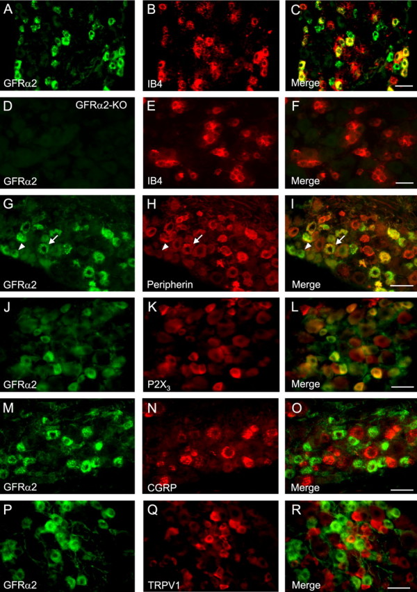
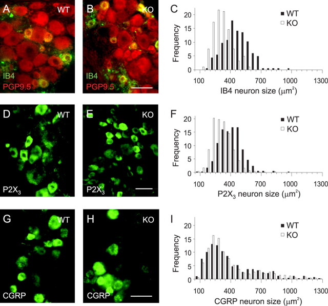
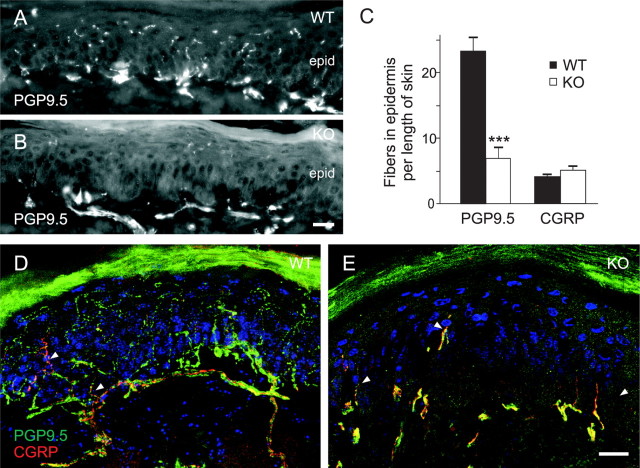
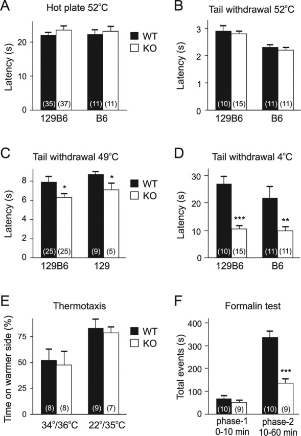
Most unmyelinated nociceptive neurons that mediate pain and temperature sensation from the skin bind isolectin B4 (IB4)-lectin and express Ret, the common signaling component of glial cell line-derived neurotrophic factor (GDNF) family. One of these factors, neurturin, is expressed in the epidermis, whereas its GDNF family receptor alpha2 (GFRalpha2) is expressed in the majority of unmyelinated Ret-positive sensory neurons. However, the physiological roles of endogenous neurturin signaling in primary sensory neurons are poorly understood. Here, we show that the vast majority (approximately 85%) of IB4 binding and P2X3 purinoreceptor-positive neurons, but virtually none of the calcitonin gene-related peptide (CGRP) or vanilloid receptor transient receptor potential vanilloid 1-positive neurons in mouse dorsal root ganglion (DRG) express GFRalpha2. In GFRalpha2 knock-out (KO) mice, the IB4-binding and P2X3-positive DRG neurons were present but reduced in size, consistent with normal number but reduced caliber of unmyelinated axons in a cutaneous nerve. Strikingly, nonpeptidergic (CGRP-negative) free nerve endings in footpad epidermis were >70% fewer in GFRalpha2-KO mice than in their wild-type littermates. In contrast, the density of CGRP-positive epidermal innervation remained unaffected. In the formalin test, the KO mice showed a normal acute response but a markedly attenuated persistent phase, indicating a deficit in inflammatory pain response. Behavioral responses of GFRalpha2-KO mice to innocuous warm and noxious heat were not blunted; the mice were actually markedly hypersensitive to noxious cold in tail immersion test. Overall, our results indicate a critical role for endogenous GFRalpha2 signaling in maintaining the size and terminal innervation of the nonpeptidergic class of cutaneous nociceptors in vivo.
Figures

Similar articles
-
Different requirements for GFRα2-signaling in three populations of cutaneous sensory neurons.PLoS One. 2014 Aug 11;9(8):e104764. doi: 10.1371/journal.pone.0104764. eCollection 2014. PLoS One. 2014. PMID: 25111710 Free PMC article.
-
Glial cell line-derived neurotrophic factor is a survival factor for isolectin B4-positive, but not vanilloid receptor 1-positive, neurons in the mouse.J Neurosci. 2002 May 15;22(10):4057-65. doi: 10.1523/JNEUROSCI.22-10-04057.2002. J Neurosci. 2002. PMID: 12019325 Free PMC article.
-
Lack of cholinergic innervation in gastric mucosa does not affect gastrin secretion or basal acid output in neurturin receptor GFRα2 deficient mice.J Physiol. 2013 Apr 15;591(8):2175-88. doi: 10.1113/jphysiol.2012.246801. Epub 2013 Jan 21. J Physiol. 2013. PMID: 23339174 Free PMC article.
-
Other neurotrophic factors: glial cell line-derived neurotrophic factor (GDNF).Microsc Res Tech. 1999 May 15-Jun 1;45(4-5):292-302. doi: 10.1002/(SICI)1097-0029(19990515/01)45:4/5<292::AID-JEMT13>3.0.CO;2-8. Microsc Res Tech. 1999. PMID: 10383122 Review.
-
Capsaicin, transient receptor potential (TRP) protein subfamilies and the particular relationship between capsaicin receptors and small primary sensory neurons.Anat Sci Int. 2006 Sep;81(3):135-55. doi: 10.1111/j.1447-073X.2006.00141.x. Anat Sci Int. 2006. PMID: 16955665 Review.
Cited by
-
Neurturin overexpression in skin enhances expression of TRPM8 in cutaneous sensory neurons and leads to behavioral sensitivity to cool and menthol.J Neurosci. 2013 Jan 30;33(5):2060-70. doi: 10.1523/JNEUROSCI.4012-12.2013. J Neurosci. 2013. PMID: 23365243 Free PMC article.
-
Analysis of nociception, sex and peripheral nerve innervation in the TMEV animal model of multiple sclerosis.Pain. 2008 Jun;136(3):293-304. doi: 10.1016/j.pain.2007.07.007. Epub 2007 Sep 4. Pain. 2008. PMID: 17766043 Free PMC article.
-
RET signaling is required for survival and normal function of nonpeptidergic nociceptors.J Neurosci. 2010 Mar 17;30(11):3983-94. doi: 10.1523/JNEUROSCI.5930-09.2010. J Neurosci. 2010. PMID: 20237269 Free PMC article.
-
Different requirements for GFRα2-signaling in three populations of cutaneous sensory neurons.PLoS One. 2014 Aug 11;9(8):e104764. doi: 10.1371/journal.pone.0104764. eCollection 2014. PLoS One. 2014. PMID: 25111710 Free PMC article.
-
Changes in Ionic Conductance Signature of Nociceptive Neurons Underlying Fabry Disease Phenotype.Front Neurol. 2017 Jul 14;8:335. doi: 10.3389/fneur.2017.00335. eCollection 2017. Front Neurol. 2017. PMID: 28769867 Free PMC article.
References
-
- Airaksinen MS, Saarma M (2002). The GDNF family: signalling, biological functions and therapeutic value. Nat Rev Neurosci 3:383–394. - PubMed
-
- Amaya F, Shimosato G, Nagano M, Ueda M, Hashimoto S, Tanaka Y, Suzuki H, Tanaka M (2004). NGF and GDNF differentially regulate TRPV1 expression that contributes to development of inflammatory thermal hyperalgesia. Eur J Neurosci 20:2303–2310. - PubMed
-
- Baloh RH, Enomoto H, Johnson EMJ, Milbrandt J (2000). The GDNF family ligands and receptors–implications for neural development. Curr Opin Neurobiol 10:103–110. - PubMed
-
- Banbury Conference on Genetic Background in Mice. (1997). Mutant mice and neuroscience: recommendations concerning genetic background. Neuron 19:755–759. - PubMed
-
- Baudet C, Mikaels A, Westphal H, Johansen J, Johansen TE, Ernfors P (2000). Positive and negative interactions of GDNF, NTN and ART in developing sensory neuron subpopulations, and their collaboration with neurotrophins. Development 127:4335–4344. - PubMed
Publication types
MeSH terms
Substances
LinkOut - more resources
Full Text Sources
Other Literature Sources
Molecular Biology Databases
Research Materials