. 2006 Feb 15;26(7):1961-70.
doi: 10.1523/JNEUROSCI.4939-05.2006.

Essential role of the main olfactory system in social recognition of major histocompatibility complex peptide ligands

Affiliations

Essential role of the main olfactory system in social recognition of major histocompatibility complex peptide ligands

Marc Spehr et al. J Neurosci. .

Abstract

Genes of the major histocompatibility complex (MHC), which play a critical role in immune recognition, influence mating preference and other social behaviors in fish, mice, and humans via chemical signals. The cellular and molecular mechanisms by which this occurs and the nature of these chemosignals remain unclear. In contrast to the widely held view that olfactory sensory neurons (OSNs) in the main olfactory epithelium (MOE) are stimulated by volatile chemosignals only, we show here that nonvolatile immune system molecules function as olfactory cues in the mammalian MOE. Using mice with targeted deletions in selected signal transduction genes (CNGA2, CNGA4), we used a combination of dye tracing, electrophysiological, Ca2+ imaging, and behavioral approaches to demonstrate that nonvolatile MHC class I peptides activate subsets of OSNs at subnanomolar concentrations in vitro and affect social preference of male mice in vivo. Both effects depend on the cyclic nucleotide-gated (CNG) channel gene CNGA2, the function of which in the nose is unique to the main population of OSNs. Disruption of the modulatory CNGA4 channel subunit reveals a profound defect in adaptation of peptide-evoked potentials in the MOE. Because sensory neurons in the vomeronasal organ (VNO) also respond to MHC peptides but do not express CNGA2, distinct mechanisms are used by the mammalian main and accessory olfactory systems for the detection of MHC peptide ligands. These results suggest a general role for MHC peptides in chemical communication even in those vertebrates that lack a functional VNO.


Figures

None
Nonvolatile molecules gain access to the mouse MOE after direct contact with the stimulus source. AF, Bright-field (A, C, E) and fluorescence images (B, D, F) showing the dissected endoturbinate system of male C57BL/6 mice. Female urine was placed on a male’s oronasal groove (ONG) (A, B, E, F) or in the anogenital region (AGR) of an estrous female, and males were allowed to freely investigate (C, D). When the urine contained the nonvolatile rhodamine dye (0.01%), rhodamine fluorescence was visible on all four endoturbinates (B, D), whereas control animals treated with rhodamine-free urine did not show such fluorescence (F). Fluorescence images in B, D, and F were taken under identical optical conditions. No significant autofluorescence was observed at this wavelength. All of these experiments used VNX mice. Roman numerals designate individual turbinates. a, Anterior; p, posterior; d, dorsal; v, ventral. Scale bars, 500 μm.
None
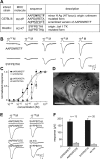
The mouse MOE detects MHC class I peptide ligands. A, Peptide ligands for prototypical representatives of MHC class I molecules used in this study. Sequences (in one-letter code) and sources were taken from Rammensee et al. (1997), which provides detailed primary references. Anchor residues defining haplotype-specific binding characteristics are highlighted in bold, and mutated residues in control peptides are underlined. In the ANPRAFDTE peptide, anchor positions 5 and 9 of the WT peptide were replaced with residues unfavorable for binding to the Db MHC molecule (Rammensee et al., 1997), whereas the overall amino acid composition remains identical. minor H Ag, Minor histocompatibility antigen; TK, tyrosine kinase. B, Examples of peptide-evoked MOE field potentials and their dose dependence (each series from a single location) to varying concentrations of either AAPDNRETF (representative of 43 recordings in 5 animals) or SYFPEITHI (representative of 38 recordings in 12 animals). Responses are produced by 500 ms pulses of peptides as indicated. C, Average dose–response plots of peak responses to AAPDNRETF (filled circles, black curve) and SYFPEITHI (open circles, red curve). The results for two control peptides, AAPDARETA (solid triangles) and SAFPEITHA (open triangles), respectively, are also shown. Smooth curves are fitted by the Hill equation, with K1/2 values and Hill coefficients of 190 pm, 0.6 (SYFPEITHI); 240 pm, 0.6 (AAPDNRETF); 5.4 nm, 0.8 (SAFPEITHA); and 8.3 nm, 1.3 (AAPDARETA). D, Medial view of the mouse endoturbinate system after dissection. Roman numerals designate individual turbinates. Sixty spatial locations of 96 spots stimulated that exhibited a field potential response to AAPDNRETF and/or SYFPEITHI at 10−10 m are shown. The other 36 locations that did not respond are included in the map in Figure 3A, which documents the entire data set. Scale bar, 1000 μm. E, F, Specificity of peptide-evoked MOE field potentials assessed by two control peptides in which the characteristic anchor residues of MHC class I ligands were replaced by alanines [i.e., AAPDARETA (8 recordings, 3 animals) and SAFPEITHA (14 recordings, 4 animals), respectively]. These peptides failed to elicit a field potential at 10−10 m (E). A scrambled version of the H2-Db ligand AAPDNRETF, ANPRAFDTE (10−10 m), and a mixture containing all amino acids (aa mix; in free form, each at 10−10 m) that constitute the SYFPEITHI peptide elicited only very small or no responses (E, F). Each pair of responses in E corresponded to the same spatial location. Error bars indicate SEM.
None
Spatial distribution of peptide-evoked field potentials on endoturbinates IIa, IIb, III, and IV. A, Map of 96 spatial locations that were stimulated sequentially with AAPDNRETF, SYFPEITHI, and 2-heptanone (each at 10−10 m). Color-coded symbols indicate whether a given location responded to none, one, two, or all three of these stimuli. Scale bar, 1000 μm. B, Examples of field potential responses registered in different locations. The numerals (1–7) refer to a given location as shown in A. Ejecting bath solution (control) onto the MOE gave no responses. C, Analysis of the data shown in A with respect to the frequency of observing a response to each of the three ligands (at 10−10 m) on a given endoturbinate (n, number of locations tested on each turbinate).
None
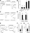
Recognition of MHC peptides by the MOE requires cAMP signaling and the CNG channel subunits A2 and A4. A, Inhibition of a peptide-induced field potential by SQ22536 (300 μm). B, Summary of the effects of pharmacological blockers on peptide-evoked potentials. Each response was normalized to its own control response obtained before application of each drug as shown in A. C, Representative peptide-induced responses from the MOE of WT and CNGA2−/− mice. Responses to 2-heptanone (10−10 m) are shown for comparison. In accord with Lin et al. (2004), we observed residual EOG responses to 2-heptanone in CNGA2−/− mice at a much higher concentration (10−4 m; n = 9) (data not shown). D, Average size of peptide-evoked MOE potentials in mice deficient for CNGA2 or CNGA4 compared with WT littermate controls. E, Representative responses to a prolonged 6 s peptide pulse in WT and CNGA4−/− MOE. Fit is a single-exponential decay with a time constant (τ) of 1.1 s. F, G, Diminished adaptation rate of peptide-evoked potentials in CNGA4−/− MOE as observed by measuring plateau/peak ratios (F) or desensitization time constants (G), respectively (means ± SEM; Student’s t test; **p < 0.001). The peptide-evoked potentials were recorded on endoturbinate III in response to SYFPEITHI (10−10 m). The number of experiments is indicated above each bar. See Results for rationale.
None
In situ Ca2+ mapping reveals distinct populations of peptide-sensitive OSNs. A, Transmitted light image of a mouse coronal MOE slice (P7). DR, Dorsal recess; S, nasal septum. A portion of the sensory epithelium delimited by the white box is shown at a higher magnification in B and C. B, Confocal fluorescence image of the fluo-4-loaded MOE acquired at rest (grayscale). C, The same region is depicted as a merged pseudocolor image of the relative increase in peptide-induced Ca2+-dependent dye fluorescence (ratio between the peak fluorescence before and after stimulation, ΔF/F). In this example, AAPDNRETF (10−9 m; green) activated three OSNs (cells 1, 2, and 4), and SYFPEITHI (10−9 m; red) activated one OSN (cell 3). In some cases, individual somata, dendrites, and dendritic knobs can be clearly distinguished. D, Time course of peptide-induced Ca2+ responses from the same cells that are shown in C. The tissue was successively stimulated with five different peptides (each at 10−9 m), followed by forskolin (20 μm). Note that cells 1 and 2 each recognized two peptides with different anchor residues. The arrows indicate the time points at which stimulus application was turned on. Scale bars: A, 200 μm; B, C, 10 μm.
None
Essential role of the main olfactory system and the CNGA2 channel subunit in the display of a social preference for disparate MHC class I peptides in male mice. A, Relative investigation time (means ± SEM) in the cotton-tip test plotted as a function of time. Bin width, 1 min. Data are based on results from animals 15–26 (preoperative) and 41–52 (see supplemental Fig. 7, available at www.jneurosci.org as supplemental material, for individual mice). For each individual animal, the investigation time per 1 min bin was measured and normalized to the maximum value. B, Preference of C57BL/6 males for urine obtained from BALB/c (gray) versus C57BL/6 (black) female mice [F(1,27) = 124.1; Student–Newman–Keuls (SNK) test; *p < 0.001]. C, Preference of C57BL/6 males for female same-strain urine supplemented with mixtures of either BALB/c (gray) or C57BL/6 (black) peptides. A preference for disparate peptides was observed in preoperative (pre-op) and VNX mice (F(1,47) = 29.5; SNK; *p < 0.01) with no significant effect of surgical VNX (SNK; p = 0.43). D, When direct physical contact is precluded (caged stimuli), C57BL/6 males do not show a preference for female same-strain urine supplemented with mixtures of BALB/c (gray) versus C57BL/6 (black) peptides (F(1,27) = 2.3; SNK; NSp = 0.16). E, Preference of BALB/c males for female same-strain urine supplemented with C57BL/6 (black) versus BALB/c (gray) peptides (F(1,23) = 10.0; SNK; *p < 0.01). F, Representative images of coronal sections through the main and accessory olfactory bulb of sham-operated and VNX mice. Sections were stained with HRP-SBA. The absence of HRP-SBA labeling in the accessory olfactory bulb of VNX animals signifies the complete removal of the VNO. GL/AOB, Glomerular layer of the accessory olfactory bulb. G, Preference of CNGA2+/0 and CNGA2−/0 males for female same-strain urine supplemented with BALB/c peptides (gray) or solvent (black). CNGA2+/0 but not CNGA2−/0 mice displayed a significant preference for disparate peptides (F(1,47) = 11.1; SNK; *p < 0.01; NSp = 0.33).

References

    1. Beauchamp GK, Yamazaki K (2003). Chemical signalling in mice. Biochem Soc Trans 31:147–151. - PubMed
    1. Boehm T, Zufall F (2006). MHC peptides and the sensory evaluation of genotype. Trends Neurosci in press. - PubMed
    1. Boehm T, Bleul CC, Schorpp M (2003). Genetic dissection of thymus development in mouse and zebrafish. Immunol Rev 195:15–27. - PubMed
    1. Boschat C, Pélofi C, Randin O, Roppolo D, Lüscher C, Broillet MC, Rodriguez I (2002). Pheromone detection mediated by a V1r vomeronasal receptor. Nat Neurosci 5:1261–1262. - PubMed
    1. Bozza T, McGann JP, Mombaerts P, Wachowiak M (2004). In vivo imaging of neuronal activity by targeted expression of a genetically encoded probe in the mouse. Neuron 42:9–21. - PubMed

Publication types