. 2006 Feb 15;26(7):1971-8.
doi: 10.1523/JNEUROSCI.2173-05.2006.

Forebrain glucocorticoid receptors modulate anxiety-associated locomotor activation and adrenal responsiveness

Affiliations

Forebrain glucocorticoid receptors modulate anxiety-associated locomotor activation and adrenal responsiveness

Maureen P Boyle et al. J Neurosci. .

Abstract

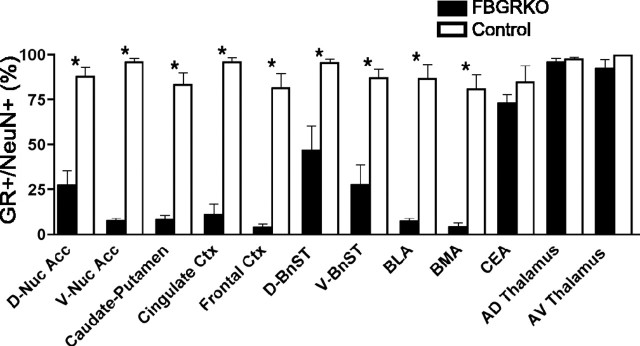
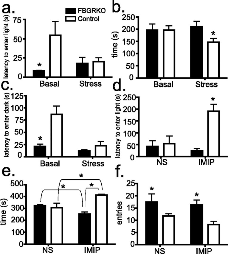
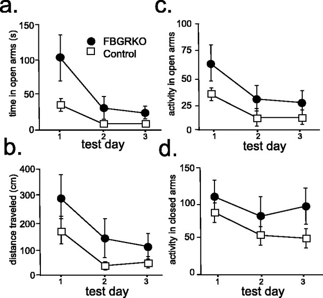
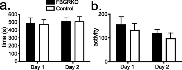
Stress potently modulates anxiety- and depression-related behaviors. In response to stressors, the hypothalamic-pituitary-adrenal (HPA) axis is activated, resulting in the release of glucocorticoids from the adrenal cortex. These hormones act peripherally to restore homeostasis but also feed back to the CNS to control the intensity and duration of the stress response. Glucocorticoids act in limbic areas of the CNS to mediate the psychological and behavioral effects of stress. In this study, we investigate the effect of forebrain-specific disruption of the glucocorticoid receptor (GR) on stress- and anxiety-related behaviors. We demonstrate that mice with disruption of forebrain GR show alterations in stress-induced locomotor activation in a number of anxiety-related behavioral paradigms. These changes are associated with alterations in stress-induced HPA axis activation and, importantly, are not attenuated by chronic treatment with the tricyclic antidepressant imipramine. These data demonstrate the importance of forebrain GR in regulation of physiological and behavioral stress reactivity and suggest that distinct pathways regulate despair- and anxiety-related behaviors.


Figures

Figure 1.
Figure 1.
Region-specific disruption of neuronal GR expression in FBGRKO mice. Neuronal GR expression was evaluated by colocalization of GR and NeuN immunoreactivity in the dorsal and ventral nucleus accumbens (D-Nuc Acc, V-Nuc Acc), caudate–putamen, cingulate cortex (Ctx), frontal cortex, dorsal and ventral bed nucleus of the stria terminalis (D-BnST, V-BnST), basolateral amygdala (BLA), basomedial amygdala (BMA), central nucleus of the amygdala (CEA), and anterodorsal (AD) and anteroventral (AV) thalamic nuclei (n = 3–4 mice per genotype, 2–4 sections per animal per region; *p < 0.05 control vs FBGRKO in specific region).
Figure 2.
Figure 2.
Altered stress reactivity in FBGRKO mice. a, In the light/dark preference test (n = 5–6), FBGRKO mice show a significantly decreased latency to enter the light compartment (*p = 0.03) compared with controls. Restraint stress significantly reduced the latency to enter the light compartment in controls but not in FBGRKO mice (p = 0.03). b, No basal differences were found between genotypes in time spent in the light compartment. After stress, control mice spent significantly less time in the light compartment (*p = 0.05). Stress had no effect on this measure in FBGRKO mice. c, In a modified version of the light/dark preference test, mice were placed in the light compartment, and the time to enter the dark compartment was measured. In the unstressed condition, FBGRKO mice show a significantly reduced latency to enter the dark chamber (*p = 0.001). Restraint stress significantly reduced the latency to enter the light compartment in controls but not in FBGRKO mice (*p = 0.003). d, The chronic mild stress associated with daily normal saline (NS) injections resulted in no difference in latency to enter the light compartment between genotypes. Treatment with imipramine (IMIP) produced a significant increase in this measure in control mice (*p = 0.03). e, Imipramine treatment (n = 4) led to a significant increase in time spent in the light compartment in control mice (p = 0.04) and a significant decrease in this measure in FBGRKO mice (p = 0.01). The net result produced a significant difference between genotypes (p < 0.0001). f, FBGRKO mice show a significantly increased (*) number of entries into the light compartment compared with controls when treated with either normal saline (p = 0.04) or imipramine (p = 0.02).
Figure 3.
Figure 3.
Anxiety-related behaviors in FBGRKO and control mice. Analysis of behavior in the elevated plus maze (n = 8–9) revealed that FBGRKO mice spend significantly more time (p = 0.03) (a) and travel significantly more distance (p = 0.05) (b) in the open arms. This change was associated with a significant increase in locomotor activity in both the open (p = 0.01) (c) and closed (p = 0.02) (d) arms of the maze.
Figure 4.
Figure 4.
No differences in open-field behavior in FBGRKO mice. a, FBGRKO and control mice took an equivalent amount of time to enter the center of the open field (quadrant 1) on both days (n = 10). Similar results were found for time to enter quadrant 2 (data not shown). b, No differences were found in the activity of FBGRKO and control mice during open-field testing as assessed by line crossing in the arena.
Figure 5.
Figure 5.
Increased hippocampal CRHR1 expression in FBGRKO mice. a, Representative sections of pseudocolored in situ hybridization images from FBGRKO and control mice with the color gradient applied from low (black) to high (red). b, Quantitative analysis of CRHR1 mRNA (n = 3–4) revealed significantly increased expression (*) in areas CA1 (p = 0.003) and the dentate gyrus (DG; p = 0.03) of the hippocampus in FBGRKO mice. CTX, Cortex.
Figure 6.
Figure 6.
HPA axis regulation after stress. a, Analysis of CORT secretion after restraint stress (n = 4–5) revealed that FBGRKO mice secrete significantly more (*) CORT after 5 and 15 min of restraint stress. Plasma CORT was also significantly higher in FBGRKO mice 60 min after the termination of restraint stress (p = 0.04), and a trend toward increased CORT secretion was found after 90 min in FBGRKO mice (†p = 0.06). b, FBGRKO mice secrete significantly more ACTH after 5 min of restraint stress compared with controls. c, FBGRKO mice secrete significantly more CORT 5, 15, and 30 min (p = 0.05) but not 60 or 90 min after mild stress. Two hours after stress, FBGRKO mice again showed increased plasma CORT compared with controls (p = 0.05). d, FBGRKO mice secrete significantly more ACTH 5 min after mild stress. e, No differences were found between genotypes in the release of CORT after ACTH stimulation. NS, Normal saline. f, Analysis of activity in the forced swim test (n = 4–6) revealed that, after restraint stress, both genotypes show increased activity, but FBGRKO mice maintain decreased activity relative to controls (p = 0.03).
Figure 7.
Figure 7.
Expression of HPA axis regulators. a, In the PVN, CRH mRNA expression is decreased after both mild (MS) (p = 0.05) and restraint stress (p = 0.006) in control but not FBGRKO mice. b, AVP expression is significantly greater (*) in the PVN of FBGRKO mice basally (p = 0.0014), after mild stress (p = 0.05), and after restraint stress (p = 0.04). c, Analysis of hippocampal MR expression by in situ hybridization in naive FBGRKO and control mice revealed no differences across genotypes. d, Analysis of MR expression after mild stress showed similar reductions in MR expression across hippocampal subfields between genotypes. e, Analysis of MR expression after restraint stress revealed significantly reduced expression (*) in hippocampal areas CA1 (p = 0.006) and the dentate gyrus (DG; p = 0.012) in the FBGRKO mice compared with stressed controls.

References

    1. Barlow DH, Campbell LA (2000). Mixed anxiety-depression and its implications for models of mood and anxiety disorders. Compr Psychiatry 41:55–60. - PubMed
    1. Belzer K, Schneier FR (2004). Comorbidity of anxiety and depressive disorders: issues in conceptualization, assessment, and treatment. J Psychiatr Pract 10:296–306. - PubMed
    1. Bjorntorp P, Rosmond R (2000). The metabolic syndrome—a neuroendocrine disorder? Br J Nutr 83:[Suppl 1], S49–S57. - PubMed
    1. Boyle MP, Brewer JA, Funatsu M, Wozniak DF, Tsien JZ, Izumi Y, Muglia LJ (2005). Acquired deficit of forebrain glucocorticoid receptor produces depression-like changes in adrenal axis regulation and behavior. Proc Natl Acad Sci USA 102:473–478. - PMC - PubMed
    1. Brewer JA, Khor B, Vogt SK, Muglia LM, Fujiwara H, Haegele KE, Sleckman BP, Muglia LJ (2003). T-cell glucocorticoid receptor is required to suppress COX-2-mediated lethal immune activation. Nat Med 9:1318–1322. - PubMed

Publication types