. 2006 Feb 15;26(7):2080-7.
doi: 10.1523/JNEUROSCI.3574-05.2006.

Presynaptic control of striatal glutamatergic neurotransmission by adenosine A1-A2A receptor heteromers

Affiliations

Presynaptic control of striatal glutamatergic neurotransmission by adenosine A1-A2A receptor heteromers

Francisco Ciruela et al. J Neurosci. .

Abstract

The functional role of heteromers of G-protein-coupled receptors is a matter of debate. In the present study, we demonstrate that heteromerization of adenosine A1 receptors (A1Rs) and A2A receptors (A2ARs) allows adenosine to exert a fine-tuning modulation of glutamatergic neurotransmission. By means of coimmunoprecipitation, bioluminescence and time-resolved fluorescence resonance energy transfer techniques, we showed the existence of A1R-A2AR heteromers in the cell surface of cotransfected cells. Immunogold detection and coimmunoprecipitation experiments indicated that A1R and A2AR are colocalized in the same striatal glutamatergic nerve terminals. Radioligand-binding experiments in cotransfected cells and rat striatum showed that a main biochemical characteristic of the A1R-A2AR heteromer is the ability of A2AR activation to reduce the affinity of the A1R for agonists. This provides a switch mechanism by which low and high concentrations of adenosine inhibit and stimulate, respectively, glutamate release. Furthermore, it is also shown that A1R-A2AR heteromers constitute a unique target for caffeine and that chronic caffeine treatment leads to modifications in the function of the A1R-A2AR heteromer that could underlie the strong tolerance to the psychomotor effects of caffeine.


Figures

Figure 1.
Figure 1.
A1R–A2AR heteromerization in HEK cells. a, Coimmunoprecipitation of A1Rs and A2ARs. HEK-293 cells transiently expressing HA-A2ARs alone (lane 1), HA-A2ARs plus Flag-A1Rs (lane 2), or Flag-A1Rs (lane 3) were washed, solubilized, and processed for immunoprecipitation using anti-Flag monoclonal antibody (2 μg/ml; IP:Flag) or anti-HA monoclonal antibody (2 μg/ml; IP:HA). Solubilized membranes (Crude) and immunoprecipitates (IP) were analyzed by SDS-PAGE and immunoblotted using rabbit anti-A2AR polyclonal antibody (1:2000) or rabbit anti-A1R polyclonal antibody (1:1000) and HRP-conjugated goat anti-rabbit IgG as a secondary antibody. These blots are representative of four different experiments with similar qualitative results. IB, Immunoblot. b, BRET saturation curve. BRET was measured in HEK-293 cells coexpressing A2AR-Rluc and A1R-YFP (▪) or A2AR-Rluc and GABABR2-YFP (▴) constructs. Cotransfections were performed with increasing amounts of plasmid DNA for the A1R-YFP construct, whereas the DNA for the A2AR-Rluc construct was maintained constant. Both fluorescence and luminescence of each sample were measured before every experiment to confirm equal expression of A2AR-Rluc while monitoring the increase of YFP expression. The saturation curve include results obtained from five independent experiments; mBU is the BRET ratio × 1000 (see Materials and Methods); error bars indicate SD of mean specific BRET ratio (mBU) values of five individual experiments grouped as a function of the amount of fluorescence of the acceptor. AU, Arbitrary unit. c, Cell surface colocalization of A1Rs and A2ARs in HEK cells. HEK cells were transfected with A2AR-HA, A1R-Flag, or both receptors simultaneously (coexpression). Nonpermeabilized cotransfected cells were immunostained with mouse anti-HA monoclonal antibody and rabbit anti-Flag polyclonal antibody. The bound primary antibodies were detected using either Alexa Fluor 488-conjugated goat anti-mouse IgG antibody or Texas Red-conjugated goat anti-rabbit. Cells were analyzed by double immunofluorescence with confocal microscopy. Superimposition of images reveals A1R–A2AR cell surface colocalization in yellow (merge). Scale bar, 10 μm.
Figure 2.
Figure 2.
A2AR-mediated modulation of A1R function in HEK cells. a, b, Modulation of [3H]R-PIA binding to A1R by A2AR activation. HEK cells were transiently cotransfected with A1R and A2AR (a) or with A1R alone (b). Membranes (0.19 mg/ml) of these cells were incubated with 0.8 nm A1R agonist [3H]R-PIA in the absence or presence of increasing concentrations of the A2AR agonist CGS21680 in 50 mm Tris-HCl containing 10 mm MgCl2 and 2 U/ml adenosine deaminase. Binding experiments were performed as described in Materials and Methods. The data were adjusted to one single binding site (dashed line) or to two binding sites (solid line). c, Modulation by A2ARs of A1R-induced Ca2+ mobilization. Transiently transfected HEK-293 cells were loaded with fura-2 AM, and the intracellular Ca2+ mobilization was determined after stimulation with R-PIA (50 nm) or CGS21680 (50 nm). The presence of CGS21680 decreases R-PIA-induced Ca2+ mobilization in the doubly transfected cells. These recordings are representative of 10 different experiments with similar qualitative results.
Figure 3.
Figure 3.
A1R–A2AR colocalization in rat striatum. a–c, Immunogold particles for the A2ARs were observed at the presynaptic level along the extrasynaptic plasma membrane (arrows) of axon terminal (B), as well as in the presynaptic active zone (crossed arrows), facing dendritic shafts (D) and spines. A2ARs were also observed at the postsynaptic level along the extrasynaptic and perisynaptic plasma membrane of dendritic shafts (D) and spines (arrowheads), establishing asymmetrical synapses with axon terminals. d, e, Similarly to A2ARs, immunogold particles for the A1R were localized along the presynaptic active zone of axon terminals (B; crossed arrows) and along the extrasynaptic plasma membrane of dendritic shafts (D) and spines (arrowheads) establishing asymmetrical synapses with axon terminals. f, g, Presynaptic colocalization of A1Rs and A2ARs. Peroxidase reaction product (immunoreactivity for A1Rs) filled axon terminals (B) establishing asymmetrical synapses with dendritic shafts (D) or spines (S), in which immunoparticles (immunoreactivity for A2ARs) were localized along the extrasynaptic plasma membrane (arrows). h, Postsynaptic colocalization of A1Rs and A2ARs. Peroxidase reaction product (immunoreactivity for A1Rs) filled dendritic shafts (D) establishing asymmetrical synapses with axon terminals (B), in which immunoparticles (immunoreactivity for A2ARs) were localized along the synaptic plasma membrane (arrowhead). Scale bar, 0.2 μm.
Figure 4.
Figure 4.
Intramembrane A1R–A2AR interaction in rat striatum. Effect of chronic caffeine treatment. Modulation of [3H]R-PIA binding to A1Rs by A2AR activation. Striatal membranes (0.25 mg/ml) of control (a) or caffeine-treated rats (b) were incubated with 0.9 nm [3H]R-PIA in the absence or presence of increasing concentrations of CGS21680 in 50 mm Tris-HCl containing 10 mm MgCl2 and 2 U/ml adenosine deaminase. Binding experiments were performed as described in Materials and Methods. The data were adjusted to one single binding site (dashed line) or to two binding sites (solid line).
Figure 5.
Figure 5.
Adenosine receptors in glutamatergic striatal nerve terminals. a, Immunocytochemical identification of A2AR (green) and A1R (blue) in the glutamatergic population of rat striatal nerve terminals (identified as vGluT-1 and vGluT-2 immunoreactive; red). In the superimposed picture of this triple-immunocytochemical labeling (merge) of this representative field, the arrows indicate the A1R/A2AR/vGluT-containing nerve terminals. b, The quantification of images of three different fields per coverslip from four experiments using different synaptosomal preparations from different animals confirmed the predominant colocalization of A1Rs and A2ARs in rat striatal glutamatergic terminals (results are means ± SEM). c, Coimmunoprecipitation of A1R and A2AR from rat striatal synaptosomes. Solubilized synaptosomes were immunoprecipitated using rabbit anti-A1R polyclonal antibody (5 μg) (lane 1), irrelevant rabbit polyclonal antibody (5 μg) (lane 2), and mouse anti-A2AR monoclonal antibody (2 μg) (lane 3). Solubilized membranes (Crude) and immunoprecipitates (lanes 1–3) were analyzed by SDS-PAGE and immunoblotted using rabbit rabbit anti-A1R antibody (1:1000) and HRP-conjugated swine anti-rabbit IgG as a secondary antibody. IB, Immunoblot.
Figure 6.
Figure 6.
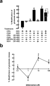
Effect of A1R and A2AR activation on evoked release of glutamate from striatal nerve terminals. Superfused synaptosomes, previously loaded with [3H]glutamate, were chemically (20 mm K+ for 30 s) stimulated twice (S1 and S2). a, CPA (100 nm) and CGS21680 (10 nm), present in S2, inhibited and facilitated, respectively, the evoked release of glutamate in a manner prevented by the A1R and A2AR antagonists DPCPX (50 nm) and SCH58261 (50 nm), respectively (present in S1 and S2). The inhibitory effect of CPA (100 nm) was also abolished when CGS21680 (10 nm) was present during S1 and S2, whereas the facilitatory effect of CGS21680 was not modified when CPA (100 nm) was present in S1 and S2. Coapplication of CPA (100 nm) and CGS (10 nm) in S2 facilitated the evoked release of glutamate. The dashed bar illustrates the arithmetic sum of the effects of 10 nm CGS21680 and 100 nm CPA, if these effects were purely additive. The results are means ± SEM of three to seven experiments. *p < 0.05 versus 0%; **p < 0.05 between bars. b, Increasing concentrations of adenosine (0.1–100 μm) produced a biphasic effect, with with low and high concentrations inhibiting and stimulating the evoked release of glutamate, respectively. The results are means ± SEM of six to eight experiments. *p < 0.05 versus 0%.

References

    1. Agnati LF, Ferré S, Lluis C, Franco R, Fuxe K (2003). Molecular mechanisms and therapeutical implications of intramembrane receptor/receptor interactions among heptahelical receptors with examples from the striatopallidal GABA neurons. Pharmacol Rev 55:509–550. - PubMed
    1. Andresen BT, Gillespie DG, Mi Z, Dubey RK, Jackson EK (1999). Role of adenosine A(1) receptors in modulating extracellular adenosine levels. J Pharmacol Exp Ther 291:76–80. - PubMed
    1. Antoniou K, Papadopoulou-Daifoti Z, Hyphantis T, Papathanasiou G, Bekris E, Marselos M, Muller M, Goldberg SR, Ferré S (2005). A detailed behavioral analysis on the motor effects of caffeine and the involvement of adenosine A1 and A2A receptors in the rat. Psychopharmacology 183:154–162. - PubMed
    1. Canals M, Marcellino D, Fanelli F, Ciruela F, de Benedetti P, Goldberg SR, Neve K, Fuxe K, Agnati LF, Woods AS, Ferré S, Lluis C, Bouvier M, Franco R (2003). Adenosine A2A-dopamine D2 receptor-receptor heteromerization: qualitative and quantitative assessment by fluorescence and bioluminescence energy transfer. J Biol Chem 278:46741–46749. - PubMed
    1. Casadó V, Canti C, Mallol J, Canela EI, Lluis C, Franco R (1990). Solubilization of A1 adenosine receptor from pig brain: characterization and evidence of the role of the cell membrane on the coexistence of high- and low-affinity states. J Neurosci Res 26:461–473. - PubMed

Publication types

MeSH terms