Comparative Study
. 2006 Feb 15;20(4):501-13.
doi: 10.1101/gad.357106.

Polycomb group-dependent Cyclin A repression in Drosophila

Affiliations
Comparative Study

Polycomb group-dependent Cyclin A repression in Drosophila

Anne-Marie Martinez et al. Genes Dev. .

Abstract

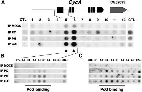
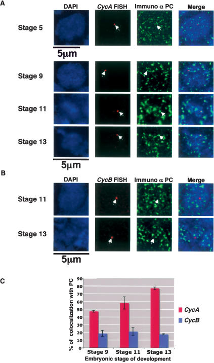
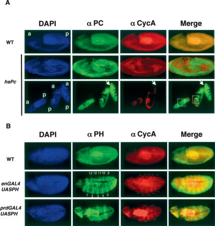
Polycomb group (PcG) and trithorax group (trxG) proteins are well known for their role in the maintenance of silent and active expression states of homeotic genes. However, PcG proteins may also be required for the control of cellular proliferation in vertebrates. In Drosophila, PcG factors act by associating with specific DNA regions termed PcG response elements (PREs). Here, we have investigated whether Drosophila cell cycle genes are directly regulated by PcG proteins through PREs. We have isolated a PRE that regulates Cyclin A (CycA) expression. This sequence is bound by the Polycomb (PC) and Polyhomeotic (PH) proteins of the PcG, and also by GAGA factor (GAF), a trxG protein that is usually found associated with PREs. This sequence causes PcG- and trxG-dependent variegation of the mini-white reporter gene in transgenic flies. The combination of FISH with PC immunostaining in embryonic cells shows that the endogenous CycA gene colocalizes with PC at foci of high PC concentration named PcG bodies. Finally, loss of function of the Pc gene and overexpression of Pc and ph trigger up-regulation and down-regulation, respectively, of CycA expression in embryos. These results demonstrate that CycA is directly regulated by PcG proteins, linking them to cell cycle control in vivo.


Figures

Figure 1.
Figure 1.
PC depletion by RNAi alters the cell cycle in proliferating S2 cells. (A) Incubation of S2 cells with double-stranded (ds) Pc RNA reduces PC expression but does not affect GAF or tubulin expression. As a control, neomycin (neo) dsRNA has no effect on PC expression. (B) Comparative FACS analysis of S2 cells after neo and Pc RNAi treatment showing altered cell cycle phasing after PC depletion. Histograms display DNA content (X-axis) and cell number (Y-axis). DNA content of neomycin and PC depleted cells is expressed in terms of G1, S, and G2 percentages.
Figure 2.
Figure 2.
Identification of a target region for PC, PH, and GAF binding within the CycA gene. (A) Slot-blot hybridization. Chromatin from Drosophila S2 cells was either mock immunoprecipitated or immunoprecipitated with anti-PC, anti-PH, or anti-GAF antibodies. One-kilobase PCR fragments from the CycA genomic region were then blotted onto a nylon membrane, and the immunopurified DNA was radiolabeled and used as a probe for hybridization (arrows indicate the signals corresponding to the strongest enrichment as compared with mock). One bound fragment is located upstream of the transcription start site (fragment 5), and the second encompasses the first exon and spreads into the neighboring intron (fragment 6). As a negative control, a fragment of 203 bp in the mini-white gene was used. As a positive control, a central region of 206 bp within the Fab-7 PRE was used. All oligonucleotide sequences are listed in the Supplemental Material. (B) The PRE products corresponding to fragments 5 and 6 were sub divided into 200-bp subfragments and subjected to hybridization using immunoprecipitated chromatin from S2 cells. PC/PH/GAF binding was detectable in four subfragments. (C) The same 200-bp subfragments were also hybridized using immunoprecipitated chromatin from stage 9–13 embryos (4–12 h after egg laying at 25°C).
Figure 3.
Figure 3.
The target region for PC/PH/GAF binding in the CycA gene causes PcG- and trxG-dependent repression of the mini-white reporter gene. (A) Eye color phenotype of two representative transgenic lines grown at 25°C. In these lines the transgene is integrated at the 28B (chromosome II) and 12E (chromosome X) cytological locations. A variegated phenotype is observed in homozygous 28B and hemizygous 12E transgenic males. (B,C) Respective effects of heterozygous PcG (PcXT109, ph410, and PcXL5) (B) and trxG (brm2)(C) mutant backgrounds on mini-white expression in the transgenic lines. Male eyes are shown.
Figure 4.
Figure 4.
PC is neither recruited at the CycA locus nor at the integration site of the transgenic PRE. Polytene chromosome immuno-FISH experiments performed on wild-type (A) and the 28B transgenic line (B) using antibodies raised against the PC protein. For each experiment, DAPI staining, immunostaining, FISH, and a merge between immunostaining and FISH are shown. In DAPI panels, the transgene insertion position is indicated by an arrow. With immunostaining, the position of the transgene is indicated by arrowheads.
Figure 5.
Figure 5.
The CycA, but not CycB, locus colocalizes with PC foci in nondividing diploid embryonic nuclei. FISH-I in whole-mount embryos. Examples of merged images of DAPI labeling (blue), PC foci (in green) after deconvolution (Supplementary Fig. S1), and the FISH probe (red) are shown for the CycA (A) and CycB (B) loci. Single slices of individual nuclei show characteristic examples of data obtained with different nuclei. (A) Cellular blastoderm stage (stage 5), early germ band elongation stage (stage 9), late germ band elongation stage (stage 11), and germ band retraction stage (stage 13) nuclei are shown for CycA. Arrows indicate cases of colocalization between CycA and PcG foci. Note the progressively larger size of the PcG body colocalizing with CycA as development progresses from stage 9 to 13. (B) Characterictic nuclei at the germ band elongation stage (stage 11) and the germ band retraction stage (stage 13) show absence of colocalization between CycB and PC foci. (C) Quantification of the percentage of colocalization between the CycA and CycB loci and PcG foci during embryonic development was performed in at least 100 nuclei per embryo. Three embryos were analyzed for each experiment.
Figure 6.
Figure 6.
CycA expression is down-regulated following Pc and ph overexpression in mitotically active embryos. (A) PC and CycA expressions are ubiquitous in wild-type stage 8 embryos (WT) and coexpression is often observed (merge). After heat-shock treatment of transgenic hsPc embryos (carrying a hsPc transgene), Pc overexpression triggers the down-regulation of CycA expression (hsPc). The middle row shows a hsPc embryo with partial ectopic PC overexpression. CycA is repressed in the PC-overexpressing domain. (Bottom row) This down-regulation is observed throughout the body plan of the affected embryos during germ band elongation stages (8–9), except in cells of the amnioserosa. Dotted squares highlight the amnioserosa region. CycA expression persists outside of the PC overexpression domains. Anterior (a) and posterior (p) parts of the embryos are indicated. The arrows point to an embryo with a strong homogeneous PC overexpression, which silenced CycA throughout the embryonic body. (B) Analysis of CycA and PH expression in wild-type and PH overexpression backgrounds during early embryogenesis. PH and CycA are ubiquitous in wild-type stage 8/9 embryos (WT). In enGAL4/UASph embryos, PH is overexpressed in the posterior part of each segment. In these PH-overexpressing stripes, CycA staining is reduced. In prdGAL4/UASph embryos, PH is overexpressed by the prd driver, and CycA staining is reduced in PH-overexpressing regions (in red). Regions of ph overexpression are highlighted in the anti-PC-labeling panel by white marks, and reported in the CycA-labeling panel.
Figure 7.
Figure 7.
Loss of function of Pc triggers ectopic expression of the CycA protein in late embryos. (AD, top panels) In wild-type (WT) embryos, CycA expression levels are very low in stage 11 (A), stage 12 (B,C), and stage 13 (D) embryos. (AD, bottom panels) At the equivalent stages in PcXL5/PcXL5 homozygous embryos, CycA is ectopically expressed in epidermal cells. (A) Stage 11 embryos, lateral view. (B) Stage 12 embryos, lateral view. (C) Stage 12 embryos, dorsal view. (D) Stage 13 embryos, lateral view.

References

    1. Bantignies F., Grimaud, C., Lavrov, S., Gabut, M., and Cavalli, G. 2003. Inheritance of Polycomb-dependent chromosomal interactions in Drosophila. Genes & Dev. 17: 2406–2420. - PMC - PubMed
    1. Beuchle D., Struhl, G., and Müller, J. 2001. Polycomb group proteins and heritable silencing of Drosophila Hox genes. Development 128: 993–1004. - PubMed
    1. Bracken A.P., Pasini, D., Capra, M., Prosperini, E., Colli, E., and Helin, K. 2003. EZH2 is downstream of the pRB–E2F pathway, essential for proliferation and amplified in cancer. EMBO J. 22: 5323–5335. - PMC - PubMed
    1. Brand A.H. and Perrimon, N. 1993. Targeted gene expression as a means of altering cell fates and generating dominant phenotypes. Development 118: 401–415. - PubMed
    1. Brehm A. and Kouzarides, T. 1999. Retinoblastoma protein meets chromatin. Trends Biochem. Sci. 24: 142–145. - PubMed

Publication types

MeSH terms

LinkOut - more resources