. 2006 Feb 21;103(8):2857-62.
doi: 10.1073/pnas.0511017103. Epub 2006 Feb 15.

A pneumococcal pilus influences virulence and host inflammatory responses

Affiliations

A pneumococcal pilus influences virulence and host inflammatory responses

M A Barocchi et al. Proc Natl Acad Sci U S A. .

Abstract

Streptococcus pneumoniae (pneumococcus) is a major cause of morbidity and mortality world-wide. The initial event in invasive pneumococcal disease is the attachment of encapsulated pneumococci to epithelial cells in the upper respiratory tract. This work provides evidence that initial bacterial adhesion and subsequent ability to cause invasive disease is enhanced by pili, long organelles able to extend beyond the polysaccharide capsule, previously unknown to exist in pneumococci. These adhesive pili-like appendages are encoded by the pneumococcal rlrA islet, present in some, but not all, clinical isolates. Introduction of the rlrA islet into an encapsulated rlrA-negative isolate allowed pilus expression, enhanced adherence to lung epithelial cells, and provided a competitive advantage upon mixed intranasal challenge of mice. Furthermore, a pilus-expressing rlrA islet-positive clinical isolate was more virulent than a nonpiliated deletion mutant, and it out-competed the mutant in murine models of colonization, pneumonia, and bacteremia. Additionally, piliated pneumococci evoked a higher TNF response during systemic infection, compared with nonpiliated derivatives, suggesting that pneumococcal pili not only contribute to adherence and virulence but also stimulate the host inflammatory response.


Conflict of interest statement

Conflict of interest statement: M.A.B., M.M., V.M., and R.R. are employees of Chiron Corporation.

Figures

Fig. 1.
Fig. 1.
Electron microscopic analysis of pneumococcal pili. (A) Negative staining of S. pneumoniae strain T4 showing abundant pili on the bacterial surface. (B) Negative staining of mutant strain T4Δ(rrgA-srtD) showing no pili. (C) Negative staining of the T4Δ(mgrA) mutant showing abundant pili. (D) Negative staining of the T4Δ(rrgA-srtD, mgrA) mutant showing no pili on the bacterial surface. (E) Immunogold labeling of T4 by using anti-RrgA. Anti-RrgA was shown to label the bacterial cell surface, suggesting that RrgA anchors the pilus structure to the cell wall. (F) Immunogold labeling of T4 with anti-RrgB (5 nm) and anti-RrgC (10 nm). Anti-RrgB was shown to decorate the entire pili. (Bar, 200 nm.) (G) High magnification of T4 pili double-labeled with anti-RrgB (5 nm) and anti-RrgC (10 nm). It shows specific labeling of a pilus tip by anti-RrgC as indicated by arrows. (Bar, 100 nm.) (H) Immunogold labeling of the deletion mutant S. pneumoniae T4Δ(rrgA-srtD) with no visible pili on the surface detectable by anti-RrgB and anti-RrgC. (Bar, 200 nm.)
Fig. 2.
Fig. 2.
Genome organization of the rlrA islet in serotype 4 strain T4 (TIGR4) and comparison with the laboratory strain R6 from available sequences. The 19F strain, ST16219F, shares a similar organization with an overall 98% sequence identity, whereas the nonencapsulated strain R6 and its progenitor D39 are pilus-islet-negative strains. A couple of insertion sequences (IS1167) flank the locus in positive strains [one of the transposases is frame-shifted (fs)], whereas an RUP element (repeat unit in pneumococcus) is identified in the pilus-islet-negative strain. The size of the locus, as well as its relative G+C content, is shown. The position of the negative regulator mgrA is indicated. Also included is the genome organization of the islets encoding pilus structures in Streptococcus agalactiae (16) and C. diphtheriae (13).
Fig. 3.
Fig. 3.
Detection of pili polymers by Western blot analysis. (A) Western blot using a 4–12% polyacrylamide gradient gel with the RrgB antiserum detects a ladder of HMW polymers in strains expressing pili (T4, T4Δ(mgrA), ST16219F, and ST16219FΔ(mgrA)), whereas the mutant strains lacking pili (T4Δ(rrgA–srtD, T4Δ(rrgA–srtD, mgrA)) and ST16219FΔ(rrgA–srtD)) have no HMW polymers. The mutant mgrA shows an increased intensity when compared with the respective wild type. (B) Western blot with the RrgB antiserum using a 4–12% gradient gel for D39 lacking the islet, the mutant D39 with the rlrA islet introduced (D39∇(rlrA–srtD), and its rlrA deletion derivative (D39∇(rlrA–srtD)∇(rlrA)).
Fig. 4.
Fig. 4.
Adherence of piliated S. pneumoniae strain D39. (A) Adherence of D39 and D39∇(rlrA–srtD), as well as D39∇(rlrA–srtD)Δ(rlrA) to monolayers of A549 lung epithelial cells. (B–D) Immunofluorescence microscopy of D39 (B), D39∇(rlrA–srtD) (C), and D39∇(rlrA–srtD)Δ(rlrA) (D) adhering to A549 lung epithelial cells. Shown are labeling of pneumococci with anti-capsular antibody (green) and visualization of epithelial F-actin with rhodamine (red).
Fig. 5.
Fig. 5.
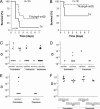
Piliated pneumococci are more virulent and outcompete nonpiliated mutants. (A–E) Intranasal challenge of C57BL/6 mice with piliated T4 and its isogenic nonpiliated deletion mutant T4Δ(rrgA-srtD). (A and B) Survival of mice after inoculation with 5 × 10⁶ cfu (high dose, A) or 5 × 10⁵ cfu (medium dose, B). Survival was analyzed by using the Kaplan–Meier log rank test. (C–E) In vivo competition infection experiments where T4 and its isogenic mutant T4Δ(rrgA–srtD) were mixed in a ratio of 1:1 before intranasal infection. The competitive index (CI) was calculated as described in Methods; each circle represents the CI for one individual mouse in each set of competition experiments. A CI below 1 indicates a competitive disadvantage of the mutant in relation to the wild-type strain. CI values <10−4 were set to 10−4. All mice were colonized. (C) CI in colonization, pneumonia, and bacteremia after high-dose challenge (n = 20). Of 20 mice, only 14 presented pneumonia (defined as bacteria recovered from the lungs), and 14 were bacteremic. We have shown previously that TIGR4 evoke an inflammatory response in murine lungs (26). (D) CI in colonization after medium dose challenge (n = 10). Of 10 mice, only 5 presented pneumonia and only 1 was bacteremic. (E) CI in colonization after low dose challenge (n = 10). Of 10 mice, only 4 presented pneumonia and none developed bacteremia. (F) CI in colonization and pneumonia after with mixed infection with wild-type D39 and its isogenic pilus islet insertion derivative D39∇(rlrA–srtD), or D39∇(rlrA–srtD)Δ(rlrA) with the rlrA gene inactivated. A CI above 1 indicates a virulence gain by the presence of the rlrA islet in D39∇(rlrA–srtD).
Fig. 6.
Fig. 6.
Role of the rlrA pilus islet in systemic host inflammatory response. Mice were challenged i.p. with high challenge dose (5 × 10⁶ to 2 × 10⁷ cfu) of T4, ST16219F, and their isogenic mutants T4Δ(rrgA–srtD), and ST16219FΔ(rrgA–srtD) and killed at 6 h after infection. (A) Bacterial outgrowth in blood after high-dose i.p. challenge. Results from individual mice are shown. Horizontal lines represent the medians, and analysis by Mann–Whitney U test gives no significant differences (P > 0.05). (B) Serum TNF response. Data are presented as means and SEMs. Statistical significance was established by Mann–Whitney U test (∗∗, P < 0.0001; ∗, P < 0.001). (C and D) TNF response for individual mice correlated to the bacteremic levels after inoculation with T4 and T4Δ(rrgA–srtD) (C) or ST16219F and ST16219FΔ(rrgA–srtD) (D).
Fig. 7.
Fig. 7.
Analysis of the IL-6 response for the same i.p. challenges as shown in Fig. 6. Bacterial growth in blood is shown in Fig. 6A. (A) Serum IL-6 response at 6 h after infection. Data are presented as means and SEMs (Mann–Whitney U test; ∗, P < 0.0001). (B) IL-6 response for individual mice correlated to the bacteremia levels after inoculation with T4 and T4Δ(rrgA–srtD).

References

    1. Bruyn G. A. W., van Furth R. Eur. J. Clin. Microbiol. Infect. Dis. 1991;10:897–910. - PubMed
    1. Ryan M. W., Antonelli P. J. Laryngoscope. 2000;110:961–964. - PubMed
    1. Cutts F. T., Zaman S. M., Enwere G., Jaffar S., Levine O. S., Okoko C., Oluwalana A., Vaughan S., Obaro A., Leach A., et al. Lancet. 2005;365:1139–1146. - PubMed
    1. Swiatlo E., Champlin F. R., Holman S. C., Wilson W. W., Watt J. M. Infect. Immun. 2002;70:412–415. - PMC - PubMed
    1. Sandgren A., Albiger B., Orihuela C., Tuomanen E., Normark S., Henriques-Normark B. J. Infect. Dis. 2005;192:791–800. - PubMed

Publication types

MeSH terms

Substances