Comparative Study
. 2006 Feb 16:7:15.
doi: 10.1186/1471-2202-7-15.

Regulation of glutamatergic signalling by PACAP in the mammalian suprachiasmatic nucleus

Affiliations
Comparative Study

Regulation of glutamatergic signalling by PACAP in the mammalian suprachiasmatic nucleus

Stephan Michel et al. BMC Neurosci. .

Abstract

Background: Previous studies indicate that light information reaches the suprachiasmatic nucleus (SCN) through a subpopulation of retinal ganglion cells that contain both glutamate and pituitary adenylyl cyclase activating peptide (PACAP). While the role of glutamate in this pathway has been well studied, the involvement of PACAP and its receptors are only beginning to be understood. Speculating that PACAP may function to modulate how neurons in the suprachiasmatic nucleus respond to glutamate, we used electrophysiological and calcium imaging tools to examine possible cellular interactions between these co-transmitters.

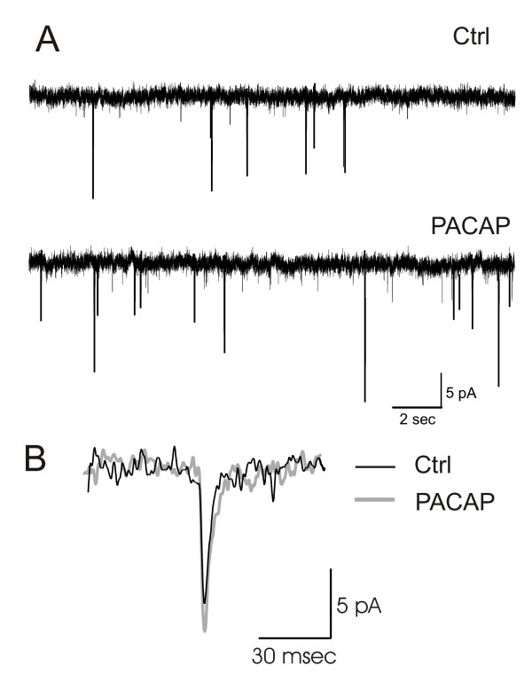
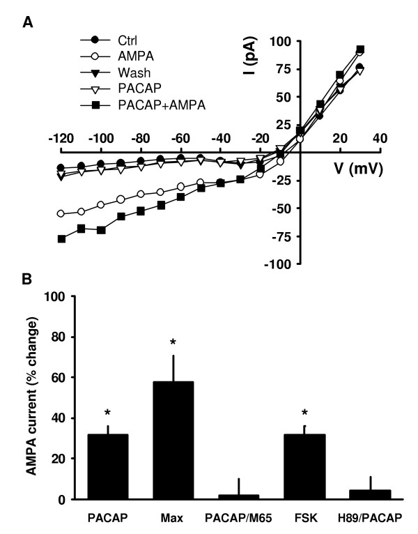
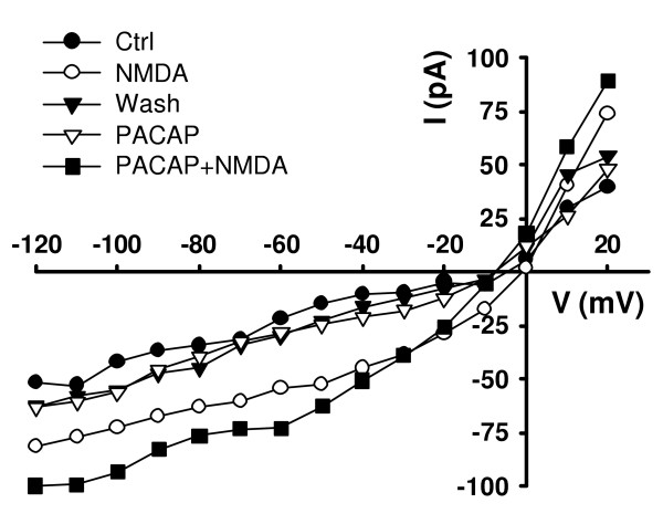
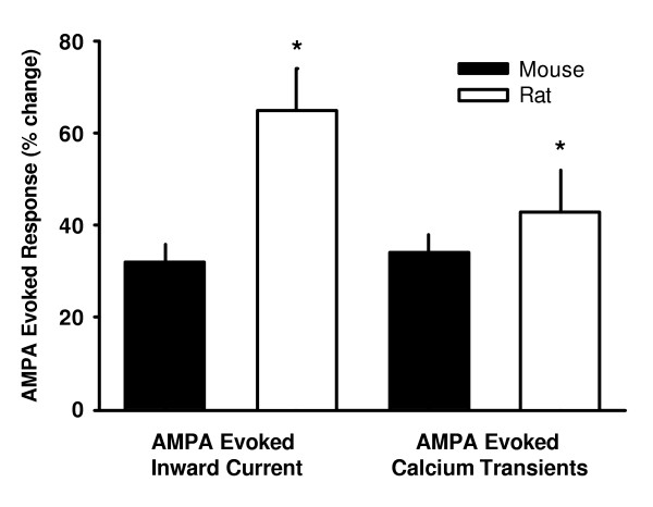
Results: Exogenous application of PACAP increased both the amplitude and frequency of spontaneous excitatory postsynaptic currents recorded from SCN neurons in a mouse brain slice preparation. PACAP also increased the magnitude of AMPA-evoked currents through a mechanism mediated by PAC1 receptors and the adenylyl cyclase-signalling cascade. This enhancement of excitatory currents was not limited to those evoked by AMPA as the magnitude of NMDA currents were also enhanced by application of PACAP. Furthermore, PACAP enhanced AMPA and NMDA evoked calcium transients while PACAP alone produced very little change in resting calcium in most mouse SCN neurons. Finally, in rat SCN neurons, exogenous PACAP enhanced AMPA evoked currents and calcium transients as well evoked robust calcium transients on its own.

Conclusion: The results reported here show that PACAP is a potent modulator of glutamatergic signalling within the SCN in the early night.


Figures

Figure 1
Figure 1
PACAP enhances the frequency and amplitude of sEPSCs recorded from mouse SCN neurons. Whole cell patch clamp recording techniques were used to measure the sEPSCs in ventral SCN neurons during the night (ZT 15–17). All experiments were carried out in presence of TTX (0.5 μM) and bicuculline (25 μM). (A) Top panel provides an example of sEPSCs recorded before and after treatment with PACAP (10 nM, 240 sec). PACAP increased the frequency (4 out of 6 neurons) and the amplitude (6 out of 6 neurons) of the sEPSCs. (B) Average sEPSC waveform recorded in an SCN neuron before and after treatment with PACAP (10 nM, 240 sec). PACAP increased the sEPSC amplitude by 20% ± 5 %.
Figure 2
Figure 2
PACAP enhanced AMPA currents in mouse SCN neurons. Whole cell patch clamp recording techniques were used to directly measure currents evoked by AMPA in ventral SCN neurons during the night (ZT 15–17). The voltage -dependence of the AMPA-evoked currents was measured by moving the membrane potential of the cell through a series of voltage -steps before, during, and after treatment with AMPA (25 μM) in the bath. (A) By itself, PACAP (10 nM, 240 sec) did not activate voltage-dependent currents, however, PACAP did increase the magnitude of AMPA-evoked currents. Top panel shows current-voltage relationships for peak current during AMPA treatment recorded using this protocol before and after treatment with PACAP (10 nM, 240 sec). (B) Histograms demonstrating that the effects of PACAP were mimicked by the PAC1 receptor agonist maxadilan and blocked by the antagonist M65. By itself, M65 did not alter the magnitude of the AMPA current (data not shown). In addition, effects of PACAP were mimicked by the AC activator FSK and blocked by the PKA inhibitor H89. By itself, H89 did not alter the magnitude of the AMPA current (data not shown). Asterisks indicate values that are significantly different than those of the AMPA-treated group (P < 0.05).
Figure 3
Figure 3
PACAP enhanced NMDA currents in mouse SCN neurons. Whole cell patch clamp recording techniques were used to directly measure currents evoked by NMDA in ventral SCN neurons during the night (ZT 15–17). The voltage -dependence of the NMDA-evoked currents was measured by moving the membrane potential of the cell through a series of voltage -steps before, during, and after treatment with NMDA (25 μM) in the bath. By itself, PACAP (10 nM, 240 sec) did not activate voltage-dependent currents, however, PACAP did increase the magnitude of NMDA-evoked currents. Current-voltage relationships for peak current during NMDA treatment recorded using this protocol before and after treatment with PACAP (10 nM).
Figure 4
Figure 4
PACAP enhanced AMPA- and NMDA-evoked Ca2+ responses in a subset of mouse SCN neurons. Optical imaging techniques were used to estimate Ca2+ responses to AMPA and NMDA in ventral SCN neurons during the night (ZT 15–17). (A) AMPA (25 μM)-induced Ca2+ transients were also enhanced by the pre-treatment with PACAP (10 nM, 240 sec). (B) NMDA (25 μM)-induced Ca2+ transients were also enhanced by the pre-treatment with PACAP (10 nM, 240 sec). (C) Histograms summarizing PACAP modulation of AMPA and NMDA-evoked Ca2+ transients in the SCN. The enhancement of AMPA-evoked Ca2+ transients was mimicked by the PAC1 receptor agonist maxadilan (Max, 100 nM, 240 sec). This data was collected in the presence of TTX. Asterisks indicate values that are significantly greater than those of the AMPA- or NMDA-treated groups (P < 0.05)
Figure 5
Figure 5
Comparison between AMPA-evoked responses in SCN neurons of rats and mice. Optical imaging techniques were used to estimate Ca2+ responses to A MPA in ventral SCN neurons during the night (ZT 15–17). Application of PACAP increased AMPA-evoked currents and Ca2+ responses in ventral SCN region of mice and rats. The left two histograms compare PACAP (10 nM, 240 sec) enhancement of AMPA (25 μM, 120 sec) induced currents in mouse and rat. The right two histograms compare PACAP (10 nM, 240 sec) enhancement of AMPA (25 μM, 120 sec) induced Ca2+ transients in mouse and rat. Pretreatment with PACAP significantly increased AMPA evoked currents and Ca2+ transients in both mice and rats. In both cases, the effects of PACAP were significantly larger in the rat. Asterisks indicate values that are significantly greater in the rat then in the mouse (P < 0.05). Data is shown as means ± SE.

Similar articles

Cited by

References

    1. Berson DM, Dunn FA, Takao M. Phototransduction by retinal gan glion cells that set the circadian clock. Science. 2002;295:1070–1073. doi: 10.1126/science.1067262. - DOI - PubMed
    1. Hattar S, Liao HW, Takao M, Berson DM, Yau KW. Melanopsin-containing retinal ganglion cells: architecture, projections, and intrinsic photosensitivity. Science. 2002;295:1065–1070. doi: 10.1126/science.1069609. - DOI - PMC - PubMed
    1. Ebling FJ. The role of glutamate in the photic regulation of the suprachiasmatic nucleus. Prog Neurobiol. 1996;50:109–132. doi: 10.1016/S0301-0082(96)00032-9. - DOI - PubMed
    1. Colwell CS, Menaker M. Excitatory Amino Acids: Their Role in Neuroendocrine Function. CRC Press; 1996. Regulation of circadian rhytms by excitatory amino acids; pp. 223–252.
    1. Colwell CS. Circadian modulation of calcium levels in cells in the suprachiasmatic nucleus. Eur J Neurosci. 2000;12:571–576. doi: 10.1046/j.1460-9568.2000.00939.x. - DOI - PMC - PubMed

Publication types

MeSH terms

Substances