. 2006 Apr 7;357(4):1058-62.
doi: 10.1016/j.jmb.2006.01.073. Epub 2006 Feb 6.

Modular enzyme design: regulation by mutually exclusive protein folding

Affiliations

Modular enzyme design: regulation by mutually exclusive protein folding

Jeung-Hoi Ha et al. J Mol Biol. .

Abstract

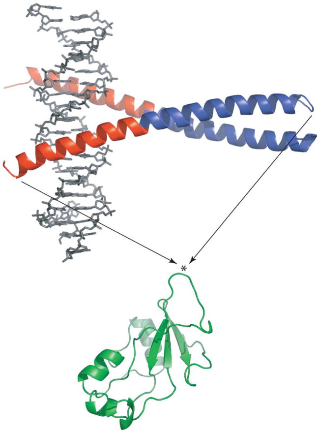
A regulatory mechanism is introduced whereupon the catalytic activity of a given enzyme is controlled by ligand binding to a receptor domain of choice. A small enzyme (barnase) and a ligand-binding polypeptide (GCN4) are fused so that a simple topological constraint prevents them from existing simultaneously in their folded states. The two domains consequently engage in a thermodynamic tug-of-war in which the more stable domain forces the less stable domain to unfold. In the absence of ligand, the barnase domain is more stable and is therefore folded and active; the GCN4 domain is substantially unstructured. DNA binding induces folding of GCN4, forcibly unfolding and inactivating the barnase domain. Barnase-GCN4 is thus a "natively unfolded" protein that uses ligand binding to switch between partially folded forms. The key characteristics of each parent protein (catalytic efficiency of barnase, DNA binding affinity and sequence specificity of GCN4) are retained in the chimera. Barnase-GCN4 thus defines a modular approach for assembling enzymes with novel sensor capabilities from a variety of catalytic and ligand binding domains.


Figures

Figure 1
Figure 1
Creation of the BG fusion protein from GCN4 (top) and Bn (bottom). The DNA binding and coiled-coil regions of GCN4 are colored red and blue, respectively. The bound DNA oligonucleotide is shown in grey. The asterisk indicates the point where GCN4 was inserted (between residues 66 and 67 of Bn). KpnI and NheI restriction sites were created to fuse the Bn and GCN4 genes. The extra nucleotides introduced Gly-Thr and Ala-Ser at the junction points. These dipeptides serve as short linkers. Images were generated by the Pymol program (DeLano Scientific).
Figure 2
Figure 2
Urea-induced denaturation of BG (filled circles) and free Bn (open squares) monitored by Trp fluorescence maximum. Lines are best fit of the data to the linear extrapolation equation. Solution conditions are 200 nM protein (monomer concentration), 25 mM Hepes (pH 7.0), 100 mM NaCl at 25 ºC. Data were collected on a Fluoromax-3 fluorometer (Jobin-Yvon/SPEX) with an excitation wavelength of 280 nm. Emission maxima were calculated using the Datamax software package (Jobin-Yvon/SPEX). BG was expressed in Escherichia coli BL21(DE3) and purified using the same protocol developed for barnase–ubiquitin. One notable difference is that BG is found completely in inclusion bodies and is thus protected from proteolysis; the yield of BG is correspondingly much higher than that of barnase–ubiquitin.
Figure 3
Figure 3
Urea dependence of the apparent dissociation constant for intermolecular complementation (Bn fragments 1–67 and 68–110). The line is meant to guide the eye only. Various concentrations of 1–67 and 68–110 Bn fragments (always present at a 1:1 ratio) were unfolded in 6 M urea then rapidly diluted to the urea concentration indicated. Refolding of the complex was monitored by shift in Trp fluorescence maximum (inset; data obtained in 0.2 M urea). Kd values were obtained by fitting fluorescence maxima to the simple 1:1 binding equation (continuous line in the inset indicates curve fit). Solution conditions are the same for Figure 2. Bn fragments 1–67 and 68–110 were prepared by digesting purified S67M mutant with CNBr.
Figure 4
Figure 4
(a) DNA binding-induced unfolding of the Bn domain of BG monitored by Trp fluorescence maximum. Samples were incubated with for 2 h with AP-1 oligonucleotide (5′-AGTGGAGATGACTCATCTCGTGC-3′) prior to measurements. (b) Inhibition of RNase activity by DNA binding. Filled and open circles designate BG incubated with AP-1 and with the non-consensus oligonucleotide 5′-CAGGGTGCTATGAACAAATGCCTCGAGCTGTTCCG T-3′, respectively. Open squares represent free Bn incubated with AP-1. Lines are best fits of the data to the simple binding equation. Fitted Kd values are ~2 nM (filled circles), 360 nM (open circles) and ~3 μM (open squares). Error bars represent standard deviations of three measurements. Samples were prepared as for (a) and assayed for RNase activity by addition of 20 μM guanylyl(3′-5′)uridine 3′-monophosphate. Substrate transesterification was monitored by absorbance at 275 nm on a Cary 100 spectrophotometer (Varian Instruments). Initial velocities were obtained from least-squares fits of the linear portion of the data. The concentration of free Bn was reduced to 60 nM to lower the initial velocity to measurable levels. Conditions are identical to those for Figure 2 except 1.4 M urea is present in all samples, and samples for enzyme assays contain 0.1 mg ml−1 bovine.
Figure 5
Figure 5
CD spectra of free Bn (broken lines) and BG (continuous lines) in the absence (grey) and presence (black) of AP-1 DNA. Protein and AP-1 concentrations were 0.50 μM and 0.94 μM, respectively. The black traces were generated by subtracting spectra of free AP-1 at the same concentration. Solution conditions are identical to those for Figure 4. Data were collected on a model 202 spectropolarimeter (Aviv Biomedical, Inc.) in a 1 cm × 1 cm cuvette. Wavelengths below 212 nm are not shown due to excessive sample absorbance.

References

    1. Radley TL, Markowska AI, Bettinger BT, Ha JH, Loh SN. Allosteric switching by mutually exclusive folding of protein domains. J Mol Biol. 2003;332:529–536. - PMC - PubMed
    1. O’Shea EK, Klemm JD, Kim PS, Alber T. X-ray structure of the GCN4 leucine zipper, a two-stranded, parallel coiled coil. Science. 1991;254:539–544. - PubMed
    1. Cranz S, Berger C, Baici A, Jelesarov I, Bosshard HR. Monomeric and dimeric bZIP transcription factor GCN4 bind a the same rate to their target DNA site. Biochemistry. 2004;43:718–727. - PubMed
    1. Zitzewitz JA, Bilsel O, Luo J, Jones BE, Matthews CR. Probing the folding mechanism of a leucine zipper peptide by stopped-flow circular dichroism spectroscopy. Biochemistry. 1995;34:12812–12819. - PubMed
    1. Zitzewitz JA, Ibarra-Molero B, Fishel DR, Terry KL, Matthews CR. Preformed secondary structure drives the association reaction of GCN4-p1, a model coiled-coil system. J Mol Biol. 2000;286:1105–1116. - PubMed

Publication types

MeSH terms

LinkOut - more resources