

Cellular and subcellular localization of PDE10A, a striatum-enriched phosphodiesterase
- PMID: 16483723
- PMCID: PMC1464838
- DOI: 10.1016/j.neuroscience.2005.12.042
Cellular and subcellular localization of PDE10A, a striatum-enriched phosphodiesterase
Abstract
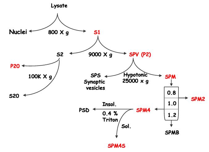
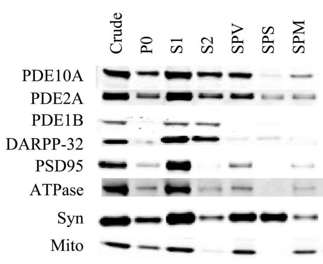
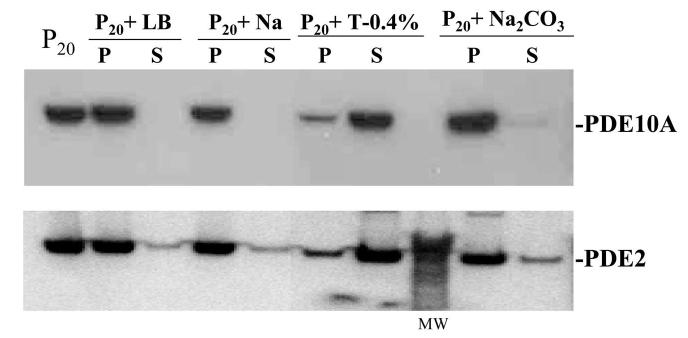
PDE10A is a recently identified phosphodiesterase that is highly expressed by the GABAergic medium spiny projection neurons of the mammalian striatum. Inhibition of PDE10A results in striatal activation and behavioral suppression, suggesting that PDE10A inhibitors represent a novel class of antipsychotic agents. In the present studies we further elucidate the localization of this enzyme in striatum of rat and cynomolgus monkey. We find by confocal microscopy that PDE10A-like immunoreactivity is excluded from each class of striatal interneuron. Thus, the enzyme is restricted to the medium spiny neurons. Subcellular fractionation indicates that PDE10A is primarily membrane bound. The protein is present in the synaptosomal fraction but is separated from the postsynaptic density upon solubilization with 0.4% Triton X-100. Immuno-electron microscopy of striatum confirms that PDE10A is most often associated with membranes in dendrites and spines. Immuno-gold particles are observed on the edge of the postsynaptic density but not within this structure. Our studies indicate that PDE10A is associated with post-synaptic membranes of the medium spiny neurons, suggesting that the specialized compartmentation of PDE10A enables the regulation of intracellular signaling from glutamatergic and dopaminergic inputs to these neurons.
Figures


References
-
- Blackstone CD, Moss SJ, Martin LJ, Levey AI, Price DL, Huganir RL. Biochemical characterization and localization of a non-N-methyl-D-aspartate glutamate receptor in rat brain. J Neurochem. 1992;58:1118–1126. - PubMed
-
- Conti M. Phosphodiesterases and cyclic nucleotide signaling in endocrine cells. Mol Endocrinol. 2000;14:1317–1327. - PubMed
-
- Conti M, Richter W, Mehats C, Livera G, Park JY, Jin C. Cyclic AMP-specific PDE4 phosphodiesterases as critical components of cyclic AMP signaling. J Biol Chem. 2003;278:5493–5496. - PubMed
-
- Coskran TM, Jakowski AB, Ramirez AD, Morton DG, Menniti FS, Schmidt CJ, Kleiman RJ, Ryan AM, Stephenson D. PDE10A: a CNS specific cGMP phosphodiesterase that is expressed in medium spiny neurons from multiple species. Society for Neuroscience 32nd annual meeting.2005.
-
- Fujishige K, Kotera J, Michibata H, Yuasa K, Takebayashi S, Okumura K, Omori K. Cloning and characterization of a novel human phosphodiesterase that hydrolyzes both cAMP and cGMP (PDE10A) J Biol Chem. 1999a;274:18438–18445. - PubMed
Publication types
MeSH terms
Substances
Grants and funding
LinkOut - more resources
Full Text Sources
Other Literature Sources
Molecular Biology Databases
