. 2006 Mar;188(5):1835-46.
doi: 10.1128/JB.188.5.1835-1846.2006.

A caulobacter crescentus extracytoplasmic function sigma factor mediating the response to oxidative stress in stationary phase

Affiliations

A caulobacter crescentus extracytoplasmic function sigma factor mediating the response to oxidative stress in stationary phase

Cristina E Alvarez-Martinez et al. J Bacteriol. 2006 Mar.

Abstract

Alternative sigma factors of the extracytoplasmic function (ECF) subfamily are important regulators of stress responses in bacteria and have been implicated in the control of homeostasis of the extracytoplasmic compartment of the cell. This work describes the characterization of sigF, encoding 1 of the 13 members of this subfamily identified in Caulobacter crescentus. A sigF-null strain was obtained and shown to be severely impaired in resistance to oxidative stress, caused by hydrogen peroxide treatment, exclusively during the stationary phase. Although sigF mRNA levels decrease in stationary-phase cells, the amount of sigma(F) protein is greatly increased at this stage, indicating a posttranscriptional control. Data obtained indicate that the FtsH protease is either directly or indirectly involved in the control of sigma(F) levels, as cells lacking this enzyme present larger amounts of the sigma factor. Increased stability of sigma(F) protein in stationary-phase cells of the parental strain and in exponential-phase cells of the ftsH-null strain is also demonstrated. Transcriptome analysis of the sigF-null strain led to the identification of eight genes regulated by sigma(F) during the stationary phase, including sodA and msrA, which are known to be involved in oxidative stress response.


Figures
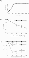
FIG. 1.
FIG. 1.
sigF mutant strain SG16 is highly sensitive to oxidative stress during the stationary phase. (A) Growth curve of NA1000 and SG16 strains. NA1000 and SG16 cells were grown in PYE complex liquid medium at 30°C, and aliquots were taken at the indicated time points to determine the OD600. (B) Survival to oxidative stress in stationary phase. Twenty-four-hour cultures of NA1000 and SG16 in PYE medium were exposed to 15 mM mM H2O2, and aliquots were taken at the indicated time points, diluted, and plated. (C) Complementation of SG16 oxidative stress sensitivity by a wild-type copy of sigF. Strains NA1000 and SG16C (sigF conditional mutant) were grown for 24 h in the absence of xylose, and then glucose (0.2%; open symbols) or xylose plus glucose (0.1% each; filled symbols) were added and cells were incubated for 150 min at 30°C. After addition of H2O2 (15 mM final concentration), aliquots of cells were taken at different times and plated. Squares, NA1000; triangles, SG16; and diamonds, SG16C. Data are representative of a minimum of three independent experiments, and the calculated standard deviations are indicated as vertical bars in each graph.
FIG. 2.
FIG. 2.
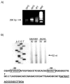
sigF is induced by extreme heat shock in an autoregulated manner. (A) RT-PCR analysis of sigF transcription after extreme heat shock during the exponential phase. Total RNA was obtained from cultures incubated at 30°C or after 15 min at 48°C. The bands correspond to amplification of the sigF transcript (arrow), detected by ethidium bromide staining of 1.5% agarose gel. −RT, without reverse transcriptase; MW, Gene Ruler molecular weight marker (MBI Fermentas). (B) Identification of the sigF transcription start site by primer extension assay. RNA samples were obtained from strains NA1000 and SG16 as described for panel A. The extension products were sized using a DNA sequencing ladder from M13mp18. The sequence below the primer extension reaction depicts the relevant features of the sigF promoter and 5′ regions. The transcription start site is indicated by an arrowhead, and the putative −35 and −10 regions are boxed. The underlined region is complementary to the oligonucleotide used as a primer in the reverse transcription reaction. The start codon suggested by TIGR annotation is denoted in italics. The data presented are representative of two independent biological experiments.
FIG. 3.
FIG. 3.
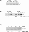
sigF is posttranscriptionally regulated in the stationary phase. (A) Levels of sigF mRNA are reduced upon entry into the stationary phase. Cell samples for RNA extraction were taken during the exponential (4 h) and stationary (12 and 24 h) phases of growth and subjected to RT-PCR analysis. The same results were obtained with two independent RNA samples. (B) The upper panel gives immunoblot analysis showing σF protein levels during different growth stages of the wild-type NA1000 strain using anti-σF polyclonal antibody. In the lower panel, the same membrane was washed and reprobed with anti-Rho polyclonal antibody as a loading control. Data from densitometry scanning of the bands are expressed below each lane as relative intensity, with time point 4 h set as 1.0.
FIG. 4.
FIG. 4.
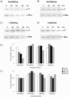
σF protein levels are affected by FtsH but not by ClpXP protease. (A) Overnight cultures of strains UJ270 (clpP) and UJ271(clpX) were grown in the presence of 0.1% xylose and 0.1% glucose, washed three times in PYE medium, and resuspended in PYE containing either 0.2% glucose (Glu) or 0.1% glucose and 0.1% xylose (Xyl). After 8 h of growth at 30°C, aliquots were taken for preparation of total protein extracts and immunoblot analysis using anti-σF antibody. (B) σF protein levels are increased in an ftsH-null mutant. Cultures from NA1000 and UJ945 (ftsH null) were grown at 30°C in PYE medium, and aliquots were taken at the indicated time points. Total protein extracts were prepared and analyzed in immunoblot assays. Results from densitometric analysis of the bands are indicated below each lane as relative intensity with respect to the less intense band. The data presented are representative of two independent biological experiments. (C) FtsH levels decrease in the stationary phase. NA1000 cells were grown in PYE liquid medium, and aliquots were taken at the indicated time points to assess FtsH levels by immunoblottng using anti-FtsH antiserum, as described in Materials and Methods. Numbers below each lane indicate FtsH levels after densitometric analysis of the corresponding band, with time point 6 h set as 1.0.
FIG. 5.
FIG. 5.
σF protein is more stable in stationary-phase cells and in an ftsH-null strain (UJ945). Overnight cultures of strains NA1000 (A and B) and UJ945 (C and D) strains were grown in PYE complex medium for 4 h (A and C, exponential phase) or 24 h (B and D, stationary [stat] phase) before addition of chloramphenicol (25 μg/ml). Aliquots were taken immediately before chloramphenicol addition (0-min samples) and at the indicated time points for preparation of total protein extracts and immunoblot analysis using anti-σF antibody. Membranes were then washed and reprobed with anti-Rho antibody as a control. Equal amounts of proteins were applied to all lanes. Relative levels of σF (E) and Rho (F) were determined by densitometric analysis of the corresponding bands, with time point zero set as 1.0. The data presented are representative of two independent biological experiments.
FIG. 6.
FIG. 6.
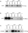
Expression of msrA, sodA, and CC3255 is down-regulated in the absence of σF. Semiquantitative RT-PCR analysis of msrA (A), sodA (B), and CC3255 (C) expression in NA1000 and SG16 strains during distinct growth phases is shown. Cultures were grown in PYE at 30°C, and aliquots were taken at the indicated time points for total RNA extraction. All reactions were performed with 19 cycles of amplification. PCR products were resolved and detected as described in Materials and Methods. The data presented are representative of two independent biological experiments.

References

    1. Alba, B. M., J. A. Leeds, C. Onufryk, C. Z. Lu, and C. A. Gross. 2002. DegS and YaeL participate sequentially in the cleavage of RseA to activate the sigma(E)-dependent extracytoplasmic stress response. Genes Dev. 16:2156-2168. - PMC - PubMed
    1. Allen, H. L. 1971. Primary productivity, chemoorganotrophy, and nutritional interactions of epiphytic algae and bacteria on macrophytes in the littoral of a lake. Ecol. Monogr. 41:97-127.
    1. Anderson, D. K., N. Ohta, J. Wu, and A. Newton. 1995. Regulation of the Caulobacter crescentus rpoN gene and function of the purified sigma 54 in flagellar gene transcription. Mol. Gen. Genet. 246:697-706. - PubMed
    1. Blaszczak, A., C. Georgopoulos, and K. Liberek. 1999. On the mechanism of FtsH-dependent degradation of the sigma 32 transcriptional regulator of Escherichia coli and the role of the Dnak chaperone machine. Mol. Microbiol. 31:157-166. - PubMed
    1. Browning, D. F., D. E. Whitworth, and D. A. Hodgson. 2003. Light-induced carotenogenesis in Myxococcus xanthus: functional characterization of the ECF sigma factor CarQ and antisigma factor CarR. Mol. Microbiol. 48:237-251. - PubMed

Publication types

MeSH terms

LinkOut - more resources