Review
. 2006 May 1;572(Pt 3):617-24.
doi: 10.1113/jphysiol.2006.105973.

Physiological roles of ATP-sensitive K+ channels in smooth muscle

Affiliations
Review

Physiological roles of ATP-sensitive K+ channels in smooth muscle

Noriyoshi Teramoto. J Physiol. .

Abstract

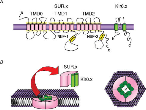
Potassium channels that are inhibited by intracellular ATP (ATP(i)) were first identified in ventricular myocytes, and are referred to as ATP-sensitive K+ channels (i.e. K(ATP) channels). Subsequently, K+ channels with similar characteristics have been demonstrated in many other tissues (pancreatic beta-cells, skeletal muscle, central neurones, smooth muscle). Approximately one decade ago, K(ATP) channels were cloned and were found to be composed of at least two subunits: an inwardly rectifying K+ channel six family (Kir6.x) that forms the ion conducting pore and a modulatory sulphonylurea receptor (SUR) that accounts for several pharmacological properties. Various types of native K(ATP) channels have been identified in a number of visceral and vascular smooth muscles in single-channel recordings. However, little attention has been paid to the molecular properties of the subunits in K(ATP) channels and it is important to determine the relative expression of K(ATP) channel components which give rise to native K(ATP) channels in smooth muscle. The aim of this review is to briefly discuss the current knowledge available for K(ATP) channels with the main interest in the molecular basis of native K(ATP) channels, and to discuss their possible linkage with physiological functions in smooth muscle.


Figures

Figure 1
Figure 1. Schematic illustration of the predicted topologies of Kir6.x and SUR.x subunits and the assembly of these subunits into a KATP channel
The transmembrane domain model proposed by Tusnady et al. (1997) is given. NBF-1 and -2 represent the two nucleotide-binding folds with Walker A and B consensus motifs. N and C indicate the N and C termini of the proteins (adapted from Cole & Clément-Chomienne, 2003). P, channel pore; Kir6.x, inwardly rectifying K+ channels; SUR.x, sulphonylurea receptors; TMD, transmembrane domain.
Figure 2
Figure 2. Effects of phenylephrine on the pinacidil-induced KATP current in smooth muscle cells dispersed from rat tail artery
Whole-cell recording, bath solution 140 mm K+ solution, pipette solution 140 mm K+ containing 5 mm EGTA. A, current trace. The vertical lines are responses to triangular ramp potential pulses of 200 mV s−1 from −120 mV to +80 mV, applied after an initial 300 ms conditioning pulse to −120 mV (see inset). Pinacidil (100 μm) caused an inward membrane current which was sustained. The current was inhibited by application of 100 μm phenylephrine. Additional application of glibenclamide suppressed the pinacidil-induced KATP current in rat tail artery. The dashed line indicates zero current. B, current–voltage relationships measured from the negative-going limb (the falling phase) of the ramp pulse. Each symbol is the same as in the current trace (A). The lines are mean membrane currents from the six ramps in each condition. C, net membrane currents. The phenylephrine-sensitive membrane current was obtained by subtraction of the membrane currents in the absence and presence of 100 μm phenylephrine when 100 μm pinacidil was present in the bath solution.
Figure 3
Figure 3. The phenylephrine-induced inhibition of KATP current was blocked by prazosin in rat tail artery
Whole-cell recording, bath solution 140 mm K+ solution, pipette solution 140 mm K+ containing 5 mm EGTA. A, current trace. The vertical lines are responses to triangular ramp potential pulses of 200 mV s−1 from −120 mV to +80 mV, applied after an initial 300 ms conditioning pulse to −120 mV (see inset). Pinacidil caused an inward membrane current which was sustained. Prazosin caused no effect on the pinacidil-induced KATP current. Similarly, additional application of phenylephrine had no effect on the pinacidil-induced KATP current in the presence of prazosin. Subsequently, application of glibenclamide suppressed the pinacidil-induced KATP current. The dashed line indicates zero current. B, current–voltage relationships measured from the negative-going limb (the falling phase) of the ramp pulse. Each symbol is the same as in the current trace (A). The lines are mean membrane currents from the six ramps in each condition. C, summary of the data. The open column indicates the current density (pA pF−1) of the pinacidil-induced KATP currents (i.e. Control). The black column shows the current density of the pinacidil-induced KATP currents in the presence of 100 μm phenylephrine (+ Pheny). The hatched column represents the current density of the pinacidil-induced KATP currents in the presence of 3 μm prazosin (+ Prazo). The grey column indicates the current density of the pinacidil-induced KATP currents in the presence of both prazosin and phenylephrine (Pheny + Prazo). *Statistically significant difference, demonstrated using a paired t test (P < 0.01). Each column represents the relative mean value with s.d. (n = 5).

Similar articles

Cited by

References

    1. Aguilar-Bryan L, Bryan J. Molecular biology of adenosine triphospate-sensitive potassium channels. Endocrine Rev. 1999;20:101–135. - PubMed
    1. Aschroft FM, Gribble FM. Correlating structure and function in ATP-sensitive K+ channels. Trends Neurosci. 1998;21:288–294. - PubMed
    1. Beech DJ, Zhang H, Nakao K, Bolton TB. K channel activation by nucleotide diphosphates and its inhibition by glibenclamide in vascular smooth muscle cells. Br J Pharmacol. 1993;110:573–582. - PMC - PubMed
    1. Beguin P, Nagashima K, Nishimura M, Gonoi T, Seino S. PKA-mediated phosphorylation of the human KATP channel: separate roles of Kir6.2 and SUR1 subunit phosphorylation. EMBO J. 1999;18:4722–4732. - PMC - PubMed
    1. Bonev AD, Nelson MT. Muscarinic inhibition of ATP-sensitive K+ channels by protein kinase C in urinary bladder smooth muscle. Am J Physiol. 1993;265:C1723–C1728. - PubMed

Publication types

MeSH terms

Substances

LinkOut - more resources