. 2006 Apr 15;572(Pt 2):335-46.
doi: 10.1113/jphysiol.2005.100776. Epub 2006 Feb 16.

Properties of ivabradine-induced block of HCN1 and HCN4 pacemaker channels

Affiliations

Properties of ivabradine-induced block of HCN1 and HCN4 pacemaker channels

A Bucchi et al. J Physiol. .

Abstract

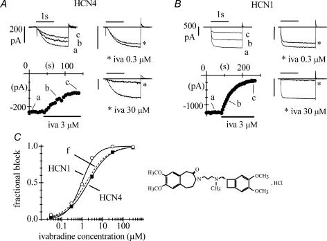
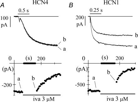
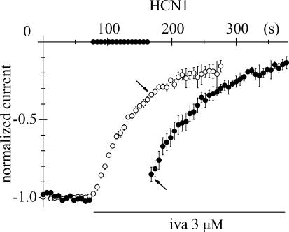
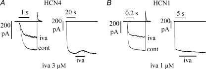
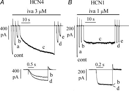
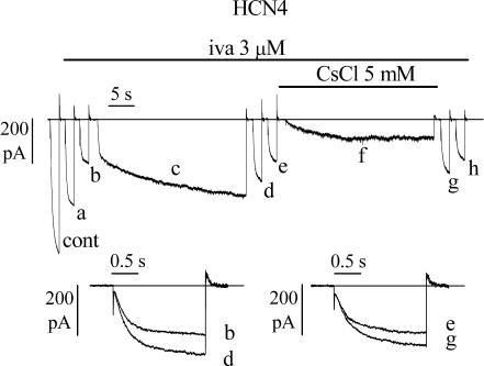
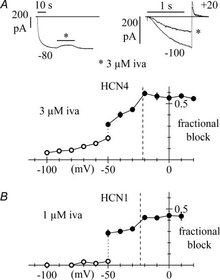
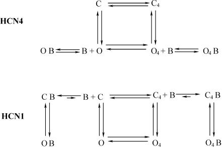
Ivabradine is a 'heart rate-reducing' agent able to slow heart rate, without complicating side-effects. Its action results from a selective and specific block of pacemaker f-channels of the cardiac sinoatrial node (SAN). Investigation has shown that block by ivabradine requires open f-channels, is use dependent, and is affected by the direction of current flow. The constitutive elements of native pacemaker channels are the hyperpolarization-activated cyclic nucleotide-gated (HCN) channels, of which four isoforms (HCN1-4) are known; in rabbit SAN tissue HCN4 is expressed strongly, and HCN1 weakly. In this study we have investigated the blocking action of ivabradine on mouse (m) HCN1 and human (h) HCN4 channels heterologously expressed in HEK 293 cells. Ivabradine blocked both channels in a dose-dependent way with half-block concentrations of 0.94 microm for mHCN1 and 2.0 microm for hHCN4. Properties of block changed substantially for the two channels. Block of hHCN4 required open channels, was strengthened by depolarization and was relieved by hyperpolarization. Block of mHCN1 did not occur, nor was it relieved, when channels were in the open state during hyperpolarization; block required channels to be either closed, or in a transitional state between open and closed configurations. The dependence of block upon current flow was limited for hHCN4, and not significant for mHCN1 channels. In summary our results indicate that ivabradine is an 'open-channel' blocker of hHCN4, and a 'closed-channel' blocker of mHCN1 channels. The mode of action of ivabradine on the two channels is discussed by implementing a simplified version of a previously developed model of f-channel kinetics.


Figures

Figure 1
Figure 1. Block of HCN4 and HCN1 channels by ivabradine
A and B, pulsing protocol for block investigation: activating/deactivating steps (−100 mV, 1.8 s; +5 mV, 0.45 s) were applied every 6 s from a holding potential of −35 mV to cells expressing either HCN4 (A) or HCN1 channels (B), and ivabradine (iva) perfused until full block development. Typical time courses of IHCN4 and IHCN1 amplitudes at −100 mV are plotted during perfusion of 3 μ m ivabradine (lower left panels in A and B). Sample current traces recorded just before (a) and during block development (b and c) for ivabradine 3 μ m are plotted in upper left panels. Sample traces recorded in control and after full block (asterisks) also shown for 0.3 and 30 μ m ivabradine in the right-hand panels in A and B. C, left, dose–response relationships of HCN4 (▪, n = 23 cells) and HCN1 (○, n = 27 cells) channel block by ivabradine (mean ± s.e.m. (most error bars are smaller than symbol size)). Each cell was exposed to one drug dose only. Fitting data points with the Hill equation resulted in half-block concentrations of 2.0 and 0.94 μ m and Hill factors of 0.8 and 1.2 for HCN4 and HCN1, respectively. C, right, ivabradine formula.
Figure 2
Figure 2. Block by ivabradine requires open HCN4, but does not require open HCN1 channels
A, time course of IHCN4 amplitude at −100 mV during a standard pulsing protocol (−100 mV, 1.8 s; +5 mV, 0.45 s). The cell was rested at −35 mV for the first 90 s of ivabradine (3 μ m) perfusion before resuming the pulsing protocol. During the period at −35 mV no current reduction was observed. B, time course of IHCN1 amplitude at −100 mV during an identical protocol. In this case, during the time spent at −35 mV a significant current reduction was indeed observed, indicating partial block development. In A and B, sample traces recorded at various times as indicated.
Figure 3
Figure 3. HCN1 block occurs preferentially when channels are cycling between open and closed states
Superimposition of the time course of block onset obtained with the protocol shown in Fig. 2B (•, mean ± s.e.m. of 6 cells) and the time course of block onset obtained with a standard activation/deactivation protocol (○, mean ± s.e.m. of 5 cells) after normalization of current amplitudes before block. The block developed during the 90 s period spent at −35 mV in the former case is less than that seen in the latter, when the pulsing protocol is not suspended (arrows), indicating that repetitive channel opening/closing cycles allow a more efficient block than having channels permanently closed.
Figure 4
Figure 4. HCN4 and HCN1 currents are blocked by ivabradine according to the protocol used to activate them
A, left, IHCN4 records in control (cont) and after full block by 3 μ m ivabradine induced by a standard activation/deactivation protocol at −100/+5 mV (as in Fig. 1); steady-state block was 60.9%. A, right, action of the same drug concentration when applied during steady-state IHCN4 activation at −100 mV; this protocol caused a block of 8.0%. B, similar experiments in an HCN1-expressing cell exposed to 1 μ m ivabradine resulted in a 50.8% block of IHCN1 with the pulsing protocol (left) and in no blocking effect when the same drug concentration was applied during steady-state IHCN1 activation at −100 mV (right).
Figure 5
Figure 5. Block of HCN4, but not HCN1 channels, is reversed by hyperpolarization
A, IHCN4 block by 3 μ m ivabradine was induced by a standard activation/deactivation protocol (−100 mV, 1.8 s; +5 mV, 1/6 Hz); records shown are: cont, control; a, 54 s, and b, 138 s after drug perfusion, the latter corresponding to steady-state block. In the continuous presence of the drug, a prolonged (40 s) step to −100 mV was then applied (trace c), following which the repetitive pulsing protocol resumed (traces d, 6 s, and e, 78 s after termination of the 40 s step). Superimposition of traces b and d (A, lower panel) shows that a partial block removal had occurred. B, in a similar experiment in an HCN1-expressing cell, a 30 s step to −100 mV was preceded and followed by standard pulsing protocols (−100 mV, 0.5 s; +5 mV, 1/6 Hz) during perfusion with ivabradine (1 μ m); records shown are: cont, control; a, 138 s, and b, 258 s after drug perfusion (corresponding to steady-state block); d, 6 s, and e, 36 s after the 30 s long step. Superimposition of traces b and d (B, lower panel) shows that the current size did not change indicating the absence of block removal.
Figure 6
Figure 6. Inward current flow contributes to hyperpolarization-induced removal of HCN4 block by ivabradine
Block by 3 μ m ivabradine was induced by a standard activation/deactivation protocol (−100/+5 mV); sample traces are shown on the left (cont, control; a, 54 s, and b, 150 s after drug application, the latter corresponding to steady-state block). A 30 s step to −100 mV was then applied in the continuous presence of the drug (trace c) and at the end of the 30 s step the repetitive pulsing protocol was resumed (traces d, 6 s, and e, 78 s after termination of the 30 s step). After reaching steady-state block for a second time (trace e), a new long (30 s) hyperpolarizing step to −100 mV was applied while simultaneously adding Cs+ (5 mm); Cs+ was washed off at the end of the new 30 s step and the repetitive pulsing protocol finally resumed (traces g, 6 s, and h, 60 s after Cs+ wash-out). Lower panels: superimposition of traces b and d, and e and g indicate that block recovery was reduced in the presence of Cs+.
Figure 7
Figure 7. Voltage-dependence of steady-state block of HCN4 and HCN1 channels by ivabradine
Data are plotted as mean ± s.e.m. averaged from 3–13 cells (A, HCN4) or 3–5 cells (B, HCN1). For both curves, at voltages equal to or more negative than −50 mV the current was activated by long steps to test potentials and the drug perfused until full block development. Fractional block was measured as the ratio between blocked and control current amplitudes (○). At voltages equal to or more positive than −50 mV, the membrane was held at the test voltage and a fixed activating voltage step to −100 mV (A: 1.2 s; B: 0.5 s) was applied repetitively (1/6 Hz). Fractional block was then measured for each test voltage as the ratio between blocked and control current at −100 mV at steady-state (•). Vertical dotted lines correspond to the mean reversal potentials measured from fully activated current–voltage relations (HCN4: −21.3 ± 1.5 mV, n = 5; HCN1: −23.5 ± 1.7 mV; n = 4). Upper panels in A represent sample IHCN4 traces for the block protocols at −80 mV (left) and +20 mV (right: asterisk indicates current at steady-state block).
Figure 8
Figure 8. Model scheme for ivabradine binding to HCN4 and HCN1 channels
The scheme is a simplification of the Altomare et al. (2001) model considering only channel states where voltage sensors are all in either the reluctant (C, O) or the willing state (C4, O4). C indicates closed channels, O open channels and B blocking molecules. Upper panel: block of HCN4. In this case the drug operates as an ‘open’ channel blocker and only interferes with open channel states. Lower panel: block of HCN1. Here free drug molecules can bind to closed, but not to open states. Further explanation in text.

Similar articles

Cited by

References

    1. Accili EA, Proenza C, Baruscotti M, DiFrancesco D. From funny current to HCN channels: 20 years of excitation. News Physiol Sci. 2002;17:32–37. - PubMed
    1. Altomare C, Bucchi A, Camatini E, Baruscotti M, Viscomi C, Moroni A, DiFrancesco D. Integrated allosteric model of voltage gating of HCN channels. J Gen Physiol. 2001;117:519–532. - PMC - PubMed
    1. Altomare C, Terragni B, Brioschi C, Milanesi R, Pagliuca C, Viscomi C, Moroni A, Baruscotti M, DiFrancesco D. Heteromeric HCN1-HCN4 channels: a comparison with native pacemaker channels from the rabbit sinoatrial node. J Physiol. 2003;549:347–359. - PMC - PubMed
    1. Baruscotti M, Bucchi A, DiFrancesco D. Physiology and pharmacology of the cardiac pacemaker (‘funny’) current. Pharmacol Ther. 2005;107:59–79. - PubMed
    1. Bois P, Bescond J, Renaudon B, Lenfant J. Mode of action of bradycardic agent, S 16257, on ionic currents of rabbit sinoatrial node cells. Br J Pharmacol. 1996;118:1051–1057. - PMC - PubMed

Publication types

MeSH terms

LinkOut - more resources