. 2006 Feb 28;103(9):3066-71.
doi: 10.1073/pnas.0600031103. Epub 2006 Feb 16.

The molecular architecture of the metalloprotease FtsH

Affiliations

The molecular architecture of the metalloprotease FtsH

Christoph Bieniossek et al. Proc Natl Acad Sci U S A. .

Abstract

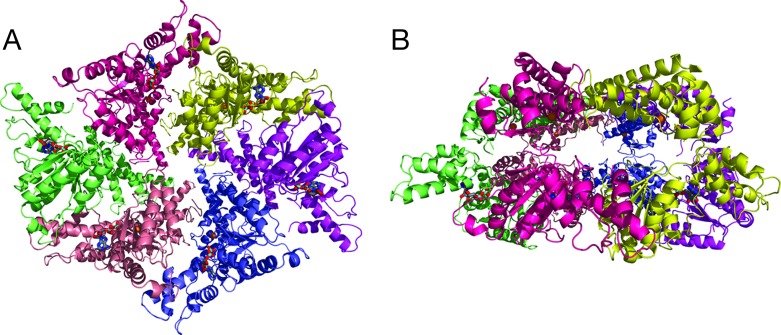
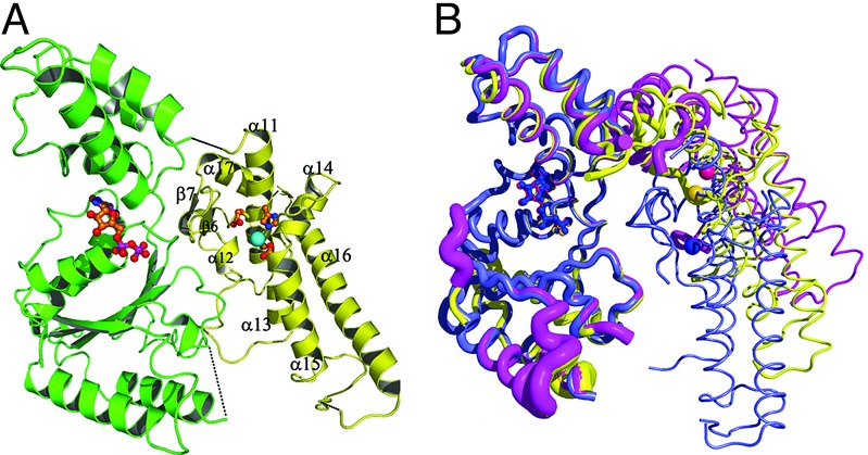
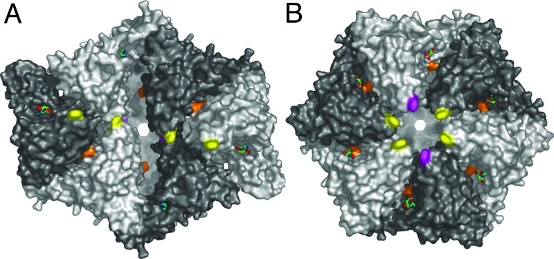
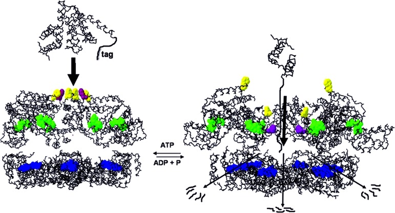
The ATP-dependent integral membrane protease FtsH is universally conserved in bacteria. Orthologs exist in chloroplasts and mitochondria, where in humans the loss of a close FtsH-homolog causes a form of spastic paraplegia. FtsH plays a crucial role in quality control by degrading unneeded or damaged membrane proteins, but it also targets soluble signaling factors like sigma(32) and lambda-CII. We report here the crystal structure of a soluble FtsH construct that is functional in caseinolytic and ATPase assays. The molecular architecture of this hexameric molecule consists of two rings where the protease domains possess an all-helical fold and form a flat hexagon that is covered by a toroid built by the AAA domains. The active site of the protease classifies FtsH as an Asp-zincin, contrary to a previous report. The different symmetries of protease and AAA rings suggest a possible translocation mechanism of the target polypeptide chain into the interior of the molecule where the proteolytic sites are located.


Conflict of interest statement

Conflict of interest statement: No conflicts declared.

Figures

Fig. 1.
Fig. 1.
Proteolytic and ATPase activity of (Δtm)FtsH. (Left) Resorufin-casein protease assay. Absorbance at 574 nm of the different samples was plotted against time. hexa, samples taken from the “early” peak of the gel-filtration purification step; mono/di, samples from the “late” peak; hexa (inhib), hexa sample incubated with 10 mM ortho-phenanthroline as inhibitor; D500A, mutant of Δtm-FtsH lacking with the true third zinc liganded mutated. The negative control (“negative”) contained no Δtm-FtsH protein. (Right) Radiogram of the [γ-32P]ATPase assay. ATP and free Pi were separated by thin-layer chromatography and are marked at the left side of the picture. Samples were taken at 5, 15, 30, 120, and 180 min. EDTA, sample incubated with 50 mM EDTA to inhibit ATPase activity.
Fig. 2.
Fig. 2.
The hexameric structure of FtsH. (A) Top view approximately down the crystallographic twofold axis from the supposed membrane side onto the AAA ring. The colors denote the individual subunits. ADP and active site residues are shown as sticks (gray, carbons; blue, nitrogens; red, oxygens; cyan, phosphorous), and the Zn2+ ions are shown as golden spheres. (B) Side view, the AAA ring is on the bottom, the protease ring on the top.
Fig. 3.
Fig. 3.
Conformation of the monomer. (A) Cartoon with secondary structure labeling. The AAA domain is shown in green, the protease domain is shown in yellow, and the zinc ion is shown in cyan. (B) Overlay of the three independent monomers of one hexamer. The AAA domain with bound ADP is at the left, and the protease domain with Zn2+ depicted as spheres is on the right. The tube thickness is proportional to the B-factors. The N-terminal part of the AAA domain was chosen as reference for the overlay. The color coding is the same as in Fig. 2.
Fig. 4.
Fig. 4.
Surface representation. (A) Top view onto AAA ring. Phe-234 residues are colored in yellow and magenta, and Arg-318 is in orange. The orientation is the same as in Fig. 2A. ADP residues are shown as sticks. Subunits are shaded alternately light and dark. (B) Modeled ideal hexameric arrangement of the AAA domains. The protease ring is in the same orientation as in A and Fig. 2A.
Fig. 5.
Fig. 5.
Schematic drawing of degradation mechanism. Side view through open-sliced molecule. The AAA ring is on top, and the protease ring is at the bottom. Phe-234 residues are colored as in Fig. 4, nucleotides are shown in green, and active site residues are in blue. After binding of an apolar recognition tag by the hydrophobic patch formed by Phe-324, ATP hydrolysis leads to the inward movement of four Phe residues, translocating the target polypeptide into the interior of the molecule followed by proteolysis.

References

    1. Sauer R. T., Bolon D. N., Burton B. M., Burton R. E., Flynn J. M., Grant R. A., Hersch G. L., Joshi S. A., Kenniston J. A., Levchenko I., et al. Cell. 2004;119:9–18. - PMC - PubMed
    1. Groll M., Bochtler M., Brandstetter H., Clausen T., Huber R. Chembiochem. 2005;6:222–256. - PubMed
    1. Ito K., Akiyama Y. Annu. Rev. Microbiol. 2005;59:211–231. - PubMed
    1. Begg K. J., Tomoyasu T., Donachie W. D., Khattar M., Niki H., Yamanaka K., Hiraga S., Ogura T. J. Bacteriol. 1992;174:2416–2417. - PMC - PubMed
    1. Jayasekera M. M., Foltin S. K., Olson E. R., Holler T. P. Arch. Biochem. Biophys. 2000;380:103–107. - PubMed

Publication types

MeSH terms

LinkOut - more resources