

Histopathologic evaluation of aneurysms treated with Guglielmi detachable coils or matrix detachable microcoils
- PMID: 16484393
- PMCID: PMC8148754
Histopathologic evaluation of aneurysms treated with Guglielmi detachable coils or matrix detachable microcoils
Abstract
Background and purpose: The purpose of this study was to evaluate the degree of organization and fibrocellular tissue development in aneurysms treated with bare platinum or biologically active microcoils.
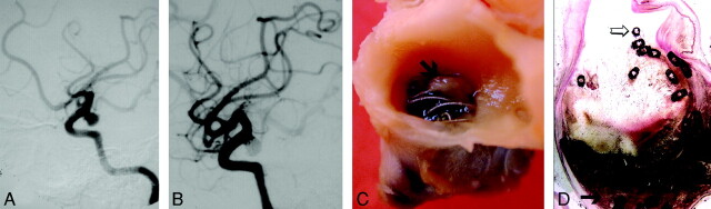
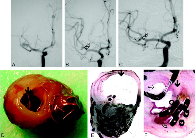
Methods: Twelve aneurysms were removed at autopsy between 1-18 days and another 2 between 2-3 months posttreatment. Four aneurysms were surgically removed between 6 months and 3 years following treatment. One aneurysm removed at 8 days and another at 6 months were treated with bioactive (Matrix) coils; the other 16 with bare platinum (Guglielmi detachable coils; GDCs). All specimens were embedded in plastic, stained with hematoxilin-eosin and elastin and examined by light microscopy.
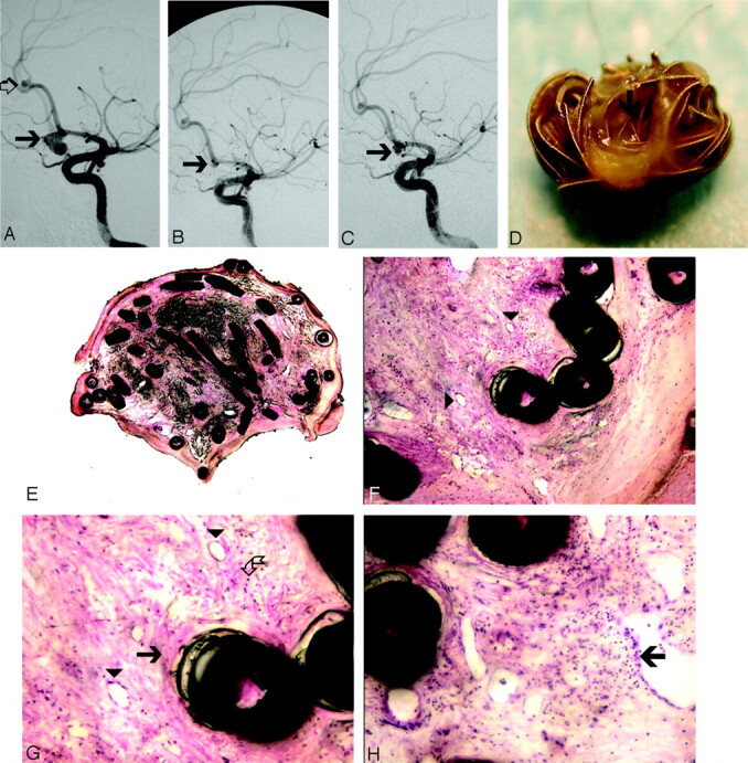
Results: All specimens removed within 3 weeks demonstrated intra-aneurysmal thrombus, without signs of organization or fibrotic tissue formation over the neck regardless of the type of coils used. In the GDC-treated aneurysms, evidence of early thrombus organization was observed within 2-3 months, and completed yet imperfect fibrocellular reaction together with residual thrombus at 2-3 years. In the Matrix-treated specimens, the aneurysm cavity was completely filled with granulation tissue corresponding to still ongoing fibrocellular reaction at 6 months, including newly formed blood vessels, smooth muscle cells, and collagen deposition without signs of residual thrombus.
Conclusions: Our results indicate that in aneurysms treated with bare platinum coils thrombus organization does not occur until late after treatment and may remain imperfect for years. In one aneurysm studied 8 days following treatment with Matrix coils, no difference was noted compared to aneurysms treated with bare platinum coils. In another aneurysm examined 6 months following packing with Matrix coils, the histologic changes support the hypothesis that the biologically active polymer may accelerate aneurysm healing.
Figures
References
-
- Molyneux AJ, Ellison DW, Morris J, et al. Histological findings in giant aneurysms treated with Guglielmi detachable coils: report of two cases with autopsy correlation. J Neurosurg 1995;83:129–32 - PubMed
-
- Stiver SI, Porter PJ, Willinsky RA, et al. Acute human histopathology of an intracranial aneurysm treated using Guglielmi detachable coils: case report and review of the literature. Neurosurgery 1998;43:1203–08 - PubMed
-
- Strother CM, Berenstein A, Vinuela F. A pitfall in the surgery of a recurrent aneurysm after coil embolization and its histological observation: technical case report. Neurosurgery 1998;42:1199–200 - PubMed
-
- Bavinzski G, Talazoglu V, Killer M, et al. Gross and microscopic histopathological findings in aneurysms of the human brain treated with Guglielmi detachable coils. J Neurosurg 1999;91:284–93 - PubMed
Publication types
MeSH terms
Substances
LinkOut - more resources
Full Text Sources
Other Literature Sources
Medical