Case Reports
. 2006 Feb;27(2):321-3.

Double spinal dural arteriovenous fistulas

Affiliations
Case Reports

Double spinal dural arteriovenous fistulas

A El-Serwi et al. AJNR Am J Neuroradiol. 2006 Feb.

Abstract

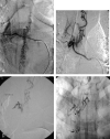
We present a patient with double spinal dural arteriovenous fistulas revealed by progressive myelopathy. Numerous dilated veins extending along the entire length of the spinal cord were found on MR imaging. Angiography showed a first spinal dural fistula at the level of T7 with descending venous drainage and a second spinal dural fistula at the level of T5 with ascending venous drainage. Both fistulas were cured by therapeutic embolization.


Figures
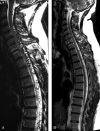
Fig 1.
Fig 1.
A, MR showing flow voids in abnormal dilated vessels extending from T12 to C4 along the spinal cord. B, Control MR 8 months after therapeutic embolization showing marked regression of the dilated vessels.
Fig 2.
Fig 2.
A, Selective angiogram of the right seventh intercostal artery showing the inferior SdAVF with a descending venous drainage. B, Selective angiogram of the right fifth intercostal artery showing the superior SdAVF with an ascending venous drainage. C, Selective angiogram of the right fourth intercostal artery after treatment of the inferior SdAVF. The venous drainage changed from exclusively ascending to both ascending and descending. The upper thoracic anterior spinal artery is visualized (arrow). D, Superselective catheterization of the meningeal feeders from the fifth intercostal artery before injection of acrylic glue. E, Casts of n-BCA at the level of T7 (arrow) and T5 (arrowhead), involving the origin of the draining veins, which enabled definite cure.
Fig 2.
Fig 2.
A, Selective angiogram of the right seventh intercostal artery showing the inferior SdAVF with a descending venous drainage. B, Selective angiogram of the right fifth intercostal artery showing the superior SdAVF with an ascending venous drainage. C, Selective angiogram of the right fourth intercostal artery after treatment of the inferior SdAVF. The venous drainage changed from exclusively ascending to both ascending and descending. The upper thoracic anterior spinal artery is visualized (arrow). D, Superselective catheterization of the meningeal feeders from the fifth intercostal artery before injection of acrylic glue. E, Casts of n-BCA at the level of T7 (arrow) and T5 (arrowhead), involving the origin of the draining veins, which enabled definite cure.

Similar articles

Cited by

References

    1. Merland JJ, Riche MC, Chiras J. Intraspinal extramedullary AV fistulae draining into the medullary veins. J Neuroradiology 1980;7:271–320 - PubMed
    1. Rosenblum B, Oldfield E, Doppman J, et al. Spinal arteriovenous malformations: a comparison of dural arteriovenous fistulae and intradural AVMs in 81 patients. J Neurosurg 1987;67:795–803 - PubMed
    1. Oldfield E, Doppman J. Spinal arteriovenous malformations. Clin Neurosurg 1988;34:161–68 - PubMed
    1. Symon L, Kuyama H, Kendall B. Dural arteriovenous malformations of the spine: clinical features and surgical results in 55 cases. J Neurosurg 1984;60:238–47 - PubMed
    1. Thron A. Spinal dural arteriovenous fistulas. Radiology 2001;41:955–60 - PubMed

Publication types