

Case Reports
The utility of DynaCT in neuroendovascular procedures
Affiliations
- PMID: 16484404
- PMCID: PMC8148775
Item in Clipboard
Case Reports
The utility of DynaCT in neuroendovascular procedures
AJNR Am J Neuroradiol.
2006 Feb.
Abstract
The authors present 3 patients who underwent neuroendovascular procedures in which DynaCT produced by a flat-panel detector facilitated management of complications. As part of a combined CT/angiography suite, DynaCT offered the major advantage of immediate detection or exclusion of intracranial complication without patient transfer. The quality of cone-volume CT-generated images produced by DynaCT was sufficient to make a diagnosis.
Figures
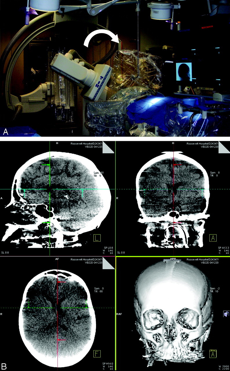
A, DynaCT acquisition demonstrating rotation of FD C-arm (curved arrow) during actual case. B, A screen shot of acquired orthogonal images immediately available to the user after acquisition and postprocessing.
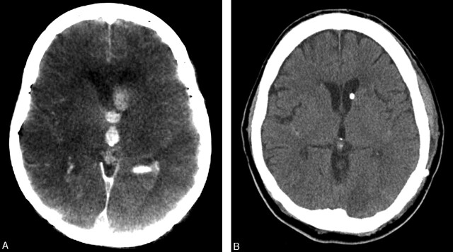
DynaCT was performed following vessel perforation (A ) and demonstrated increased ventriculomegaly compared with preoperative CT scan (B).
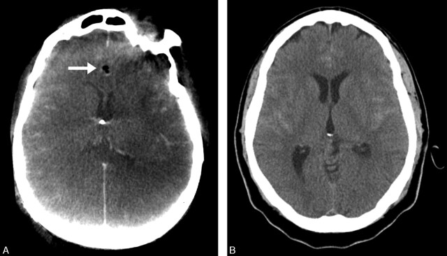
This patient experienced cardiac arrest and aneurysm rehemorrhage was suspected. After emergent ventriculostomy placement, however, DynaCT (A) demonstrated no new intracranial hemorrhage, which indicates a primary cardiac event. Note good catheter placement and interhemispheric air (arrow). Preoperative CT scan (B) is shown for comparison.
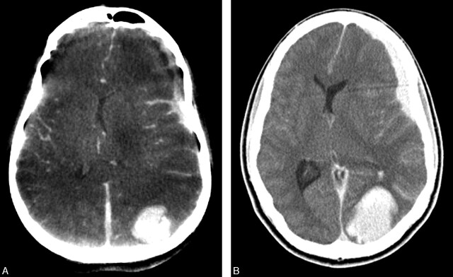
DynaCT (A) demonstrated unexpected subdural hematoma and occipitoparietal intraparenchymal hemorrhage. A conventional CT scan (B) was later obtained before patient transfer to the operating room, which revealed increased hemorrhage and mass effect.
References
-
- Horowitz MB, Crammond D, Balzer J, et al. Aneurysm rupture during endovascular coiling: effects on cerebral transit time and neurophysiologic monitoring and the benefits of early ventriculostomy: case report. Minim Invasive Neurosurg 2003;46:300–305 - PubMed
-
- Levy E, Koebbe CJ, Horowitz MB, et al. Rupture of intracranial aneurysms during endovascular coiling: management and outcomes. Neurosurgery 2001;49:807–11; discussion 811–13 - PubMed
-
- Akpek S, Brunner T, Benndorf G, et al. Three-dimensional imaging and cone beam volume CT in C-arm angiography with flat panel detector. Diagn Interv Radiol 2005;11:10–13 - PubMed
-
- Furlan A, Higashida R, Wechsler L, et al. Intra-arterial prourokinase for acute ischemic stroke: the PROACT II study: a randomized controlled trial: Prolyse in Acute Cerebral Thromboembolism. JAMA 1999;282:2003–11 - PubMed