

Case Reports
Percutaneous sacroplasty using CT fluoroscopy
Affiliations
- PMID: 16484410
- PMCID: PMC8148807
Item in Clipboard
Case Reports
Percutaneous sacroplasty using CT fluoroscopy
AJNR Am J Neuroradiol.
2006 Feb.
Abstract
Sacral insufficiency fractures frequently cause significant pain and limit activities of daily living in patients with osteoporosis. Percutaneous vertebroplasty is a common procedure to alleviate the pain associated with thoracic and lumbar vertebral compression fractures. The sacral percutaneous vertebroplasty procedure (sacroplasty) has recently been introduced as an alternative to medical management of osteoporotic sacral insufficiency fractures. We describe our CT fluoroscopy technique in performing percutaneous sacroplasty.
Figures
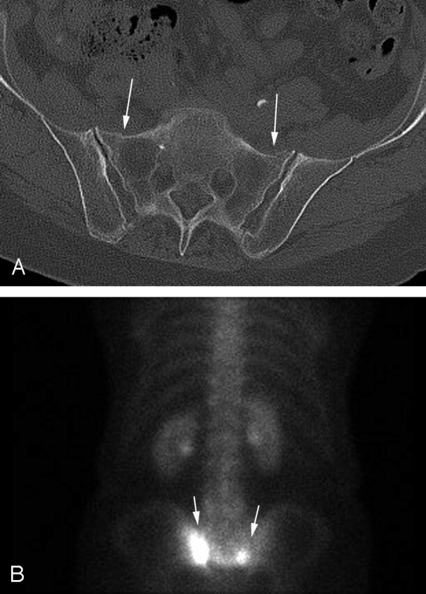
A, Axial pelvic CT demonstrates subtle bilateral sacral insufficiency fractures (arrows). B, Bone scintigraphy examination demonstrates increased radiotracer uptake (arrows) in the sacral insufficiency fractures.
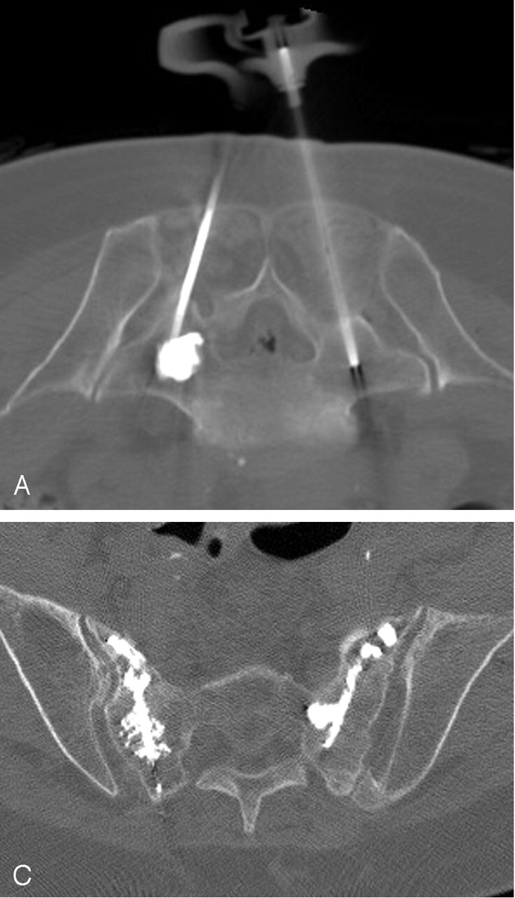
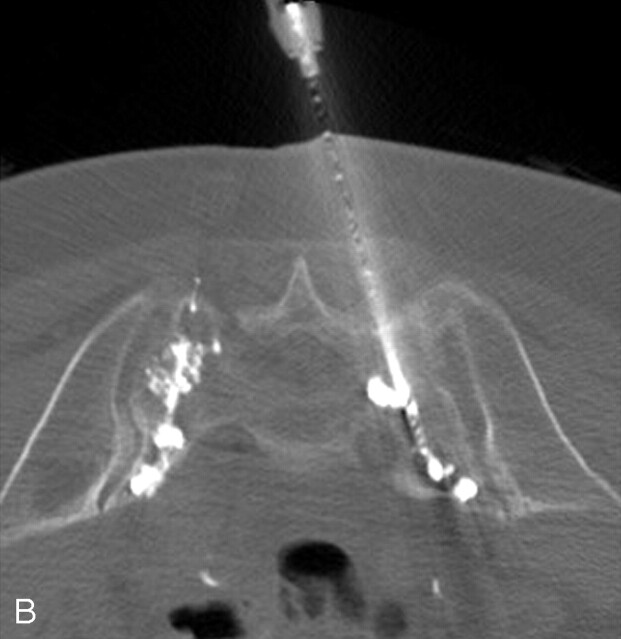
A, Axial CT fluoroscopic image demonstrates clear visualization of the injected cement in the left S1 level and the second needle on the right. B, CT fluoroscopic image during injection of the right S1 level demonstrates the cement tracts bilaterally. C, Postprocedure sacral CT demonstrates excellent cement infiltration within the bilateral sacral ala.
A, Axial CT fluoroscopic image demonstrates clear visualization of the injected cement in the left S1 level and the second needle on the right. B, CT fluoroscopic image during injection of the right S1 level demonstrates the cement tracts bilaterally. C, Postprocedure sacral CT demonstrates excellent cement infiltration within the bilateral sacral ala.
Similar articles
-
Multicenter study to assess the efficacy and safety of sacroplasty in patients with osteoporotic sacral insufficiency fractures or pathologic sacral lesions.J Neurointerv Surg. 2013 Sep 1;5(5):461-6. doi: 10.1136/neurintsurg-2012-010347. Epub 2012 Jun 8. J Neurointerv Surg. 2013. PMID: 22684691
-
[Percutaneous sacroplasty].Rev Med Suisse. 2006 Jul 12;2(73):1747-51. Rev Med Suisse. 2006. PMID: 16895111 Review. French.
-
Sacroplasty by CT and fluoroscopic guidance: is the procedure right for your patient?AJNR Am J Neuroradiol. 2007 Jan;28(1):38-41. AJNR Am J Neuroradiol. 2007. PMID: 17213421 Free PMC article.
-
Interpedicular approach in percutaneous sacroplasty for treatment of sacral vertebral body pathologic fractures.Cardiovasc Intervent Radiol. 2011 Feb;34 Suppl 2:S282-7. doi: 10.1007/s00270-010-9855-5. Epub 2010 Apr 29. Cardiovasc Intervent Radiol. 2011. PMID: 20429005
-
[Osteoporotic sacral fracture--a painful condition, easy to miss. Sacroplasty a new treatment with quick pain relief].Lakartidningen. 2010 Feb 3-9;107(5):251-4. Lakartidningen. 2010. PMID: 20297565 Review. Swedish. No abstract available.
Cited by
-
CT fluoroscopy-guided percutaneous vertebroplasty in spinal malignancy: technical results, PMMA leakages, and complications in 202 patients.Skeletal Radiol. 2012 Nov;41(11):1391-400. doi: 10.1007/s00256-012-1365-x. Skeletal Radiol. 2012. PMID: 22286549
-
Imaging and treatment of sacral insufficiency fractures.AJNR Am J Neuroradiol. 2010 Feb;31(2):201-10. doi: 10.3174/ajnr.A1666. Epub 2009 Sep 17. AJNR Am J Neuroradiol. 2010. PMID: 19762463 Free PMC article. Review.
-
Percutaneous sacroplasty with the use of C-arm flat-panel detector CT: technical feasibility and clinical outcome.Skeletal Radiol. 2011 Apr;40(4):453-60. doi: 10.1007/s00256-010-0959-4. Epub 2010 May 15. Skeletal Radiol. 2011. PMID: 20473493
-
Clinical outcomes of sacroplasty in sacral insufficiency fractures: a review of the literature.Eur Spine J. 2009 Sep;18(9):1266-71. doi: 10.1007/s00586-009-1048-z. Epub 2009 Jun 6. Eur Spine J. 2009. PMID: 19504130 Free PMC article. Review.
-
Therapeutic considerations of percutaneous sacroplasty for the sacral insufficiency fracture.J Korean Neurosurg Soc. 2010 Jan;47(1):58-63. doi: 10.3340/jkns.2010.47.1.58. Epub 2010 Jan 31. J Korean Neurosurg Soc. 2010. PMID: 20157381 Free PMC article.
References
-
- Jensen ME. Percutaneous vertebroplasty: a new therapy for the treatment of painful vertebral body compression fractures. Appl Radiol 2000;6: 7 –11
-
- Deramond H, Depriester C, Galibert P, et al. Percutaneous vertebroplasty with polymethylmethacrylate: technique, indications, and results. Radiol Clin North Am 1998;36:3:533–46 - PubMed
-
- Garant M. Sacroplasty: a new treatment for sacral insufficiency fracture. J Vasc Interv Radiol 2002;13:1265–67 - PubMed
Publication types
MeSH terms
Substances
LinkOut - more resources
Full Text Sources
Medical