

The study of cerebral hemodynamics in the hyperacute stage of fat embolism induced by triolein emulsion
- PMID: 16484418
- PMCID: PMC8148804
The study of cerebral hemodynamics in the hyperacute stage of fat embolism induced by triolein emulsion
Abstract
Purpose: The purpose of this study was to evaluate the cerebral hemodynamic change in the hyperacute stage of cerebral fat embolism induced by triolein emulsion, by using MR perfusion imaging in cat brains.
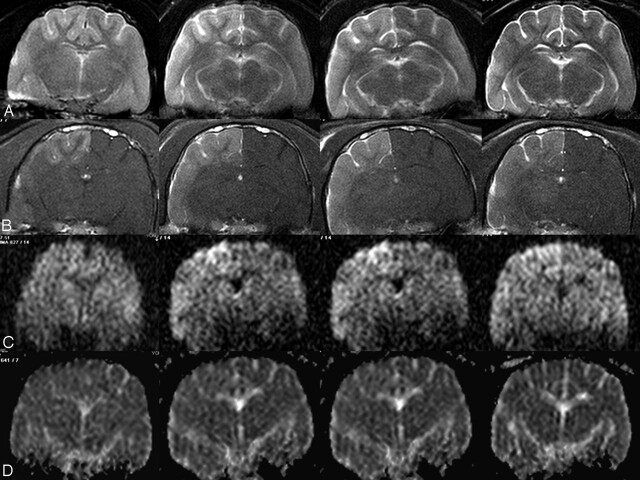
Methods: By using the femoral arterial approach, the internal carotid arteries of 14 cats were infused with an emulsion of triolein 0.05 mL. T2-weighted (T2WI), diffusion-weighted (DWI), apparent diffusion coefficient (ADC) map, perfusion-weighted (PWI), and gadolinium-enhanced T1-weighted (Gd-T1WI) images were obtained serially at 30 minutes and 2, 4, and 6 hours after infusion. The MR images were evaluated qualitatively and quantitatively. Qualitative evaluation was performed by assessing the signal intensity of the serial MR images. Quantitative assessment was performed by comparing the signal-intensity ratio (SIR) of the lesions to the contralateral normal side calculated on T2WIs, Gd-T1WIs, DWIs, and ADC maps at each acquisition time and by comparing the relative cerebral blood volume (rCBV), cerebral blood flow (CBF), and mean transit times (MTT) of the lesions to the contralateral normal side calculated on PWI.
Results: In the qualitative evaluation of the MR images, the lesions showed hyperintensity on T2WIs, enhancement on the Gd-T1WIs, and isointensity on DWIs and the ADC maps. In the quantitative studies, SIRs on the Gd-T1WIs, DWIs, and ADC maps peaked at 2 hours after infusion. The SIRs on the T2WIs peaked at 4 hours after infusion and decreased thereafter. On PWIs, the rCBV, rCBF, and MTT of the lesion showed no significant difference from the contralateral normal side (P = .09, .30, and .13, respectively) and showed no significant change of time course (P = .17, .31, and .66, respectively).
Conclusion: The embolized lesions induced by triolein emulsion showed no significant difference in cerebral hemodynamic parameters from those on the contralateral normal side. The result may suggest that consideration of the hemodynamic factor of embolized lesions is not necessary in further studies of the blood-brain barrier with triolein emulsion.
Figures
Similar articles
-
Magnetic resonance imaging and histologic findings of experimental cerebral fat embolism.Invest Radiol. 2003 Oct;38(10):625-34. doi: 10.1097/01.rli.0000077055.48406.e2. Invest Radiol. 2003. PMID: 14501490
-
Prominent hypointense veins on susceptibility weighted image in the cat brain with acute infarction: DWI, SWI, and PWI.Acta Radiol. 2014 Oct;55(8):1008-14. doi: 10.1177/0284185113508181. Epub 2013 Oct 17. Acta Radiol. 2014. PMID: 24136983
-
Experimental cerebral fat embolism: embolic effects of triolein and oleic acid depicted by MR imaging and electron microscopy.AJNR Am J Neuroradiol. 2002 Oct;23(9):1516-23. AJNR Am J Neuroradiol. 2002. PMID: 12372741 Free PMC article.
-
Diffusion-weighted MR of the brain: methodology and clinical application.Radiol Med. 2005 Mar;109(3):155-97. Radiol Med. 2005. PMID: 15775887 Review. English, Italian.
-
Imaging biomarkers guided anti-angiogenic therapy for malignant gliomas.Neuroimage Clin. 2018 Jul 5;20:51-60. doi: 10.1016/j.nicl.2018.07.001. eCollection 2018. Neuroimage Clin. 2018. PMID: 30069427 Free PMC article. Review.
Cited by
-
Triolein emulsion infusion into the hepatic artery increases vascular permeability to doxorubicin in rabbit liver.World J Gastroenterol. 2021 Jan 14;27(2):152-161. doi: 10.3748/wjg.v27.i2.152. World J Gastroenterol. 2021. PMID: 33510556 Free PMC article.
-
The minimum percentage of triolein emulsion for studying cerebral vascular permeability with least brain edema.Iran J Radiol. 2014 Sep 23;11(4):e14887. doi: 10.5812/iranjradiol.14887. eCollection 2014 Dec. Iran J Radiol. 2014. PMID: 25780547 Free PMC article.
-
Unsaturated free fatty acid emulsion infusion into carotid artery enhances drug delivery to the rat brain.Brain Behav. 2023 Jun;13(6):e2994. doi: 10.1002/brb3.2994. Epub 2023 May 22. Brain Behav. 2023. PMID: 37218399 Free PMC article.
-
Temporal profiles of aquaporin 4 expression and astrocyte response in the process of brain damage in fat embolism model in rats.J Anesth. 2010 Apr;24(2):225-33. doi: 10.1007/s00540-009-0831-7. Epub 2010 Jan 29. J Anesth. 2010. PMID: 20111877
-
Hemorrhage in cerebral fat embolisms in a cat model using triolein dependent on the physical properties of triolein.Jpn J Radiol. 2014 Jan;32(1):30-7. doi: 10.1007/s11604-013-0265-x. Epub 2013 Nov 28. Jpn J Radiol. 2014. PMID: 24288099
References
-
- Sevitt S. The significance and pathology of fat embolism. Ann Clin Res 1977;9:173–80 - PubMed
-
- Takahashi M, Suzuki R, Osakabe Y, et al. Magnetic resonance imaging findings in cerebral fat embolism: correlation with clinical manifestations. J Trauma 1999;46:324–27 - PubMed
-
- Kim HJ, Lee CH, Lee SH, et al. Magnetic resonance imaging and histologic findings of experimental cerebral fat embolism. Invest Radiol 2003;38:625–34 - PubMed
-
- Robinson EF, Soloway HB. Experimental cerebral fat embolism: distribution of radioactive triolein following internal carotid introduction. Arch Neurol 1971;24:419–22 - PubMed
Publication types
MeSH terms
Substances
LinkOut - more resources
Full Text Sources
Miscellaneous