

Case Reports
Cerebral alveolar echinoccosis mimicking primary brain tumor
Affiliations
- PMID: 16484422
- PMCID: PMC8148798
Item in Clipboard
Case Reports
Cerebral alveolar echinoccosis mimicking primary brain tumor
AJNR Am J Neuroradiol.
2006 Feb.
Abstract
We present a case of cerebral infestation by Echinococcosis multilocularis mimicking an infiltrative primary brain tumor. A heavily calcified mass invading the midbrain enhanced in a cauliflower-like fashion with small peripheral nodules present on MR imaging. Perfusion-weighted MR imaging revealed low relative cerebral blood volume within the calcified lesion and peripheral hyperemia. Single-voxel proton MR spectroscopy with an echo time of 135 milliseconds was normal.
Figures

Cranial CT shows left thalamic calcified mass (arrow) with profound edema.

Axial T2-weighted imaging (TR/TE, 3080/93 milliseconds) shows calcified mass with microcysts extending between the left thalamus (A) and the midbrain (B). Axial T1-weighted postcontrast imaging (TR/TE, 650/9 milliseconds) reveals irregular enhancement of the nodules in a cauliflower-like pattern (C) and an enhancing rim around the mass (D).
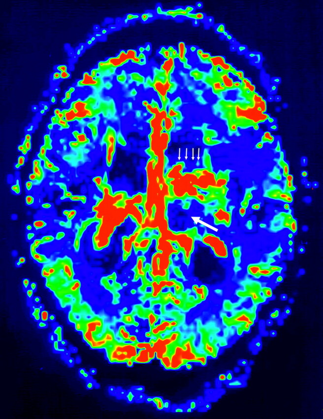
rCBV map of perfusion-weighted MR imaging (EPI [TR/TE, 1430/46 milliseconds]; NEX, 1; section thickness, 5 mm; intersection gap,10 mm; mtx, 128 ×128) demonstrates low rCBV within the lesion (arrow) located at left thalamus with higher values peripherally (thin arrows).

Multiple cysts (arrows) in the brain parenchyma surrounded by necrosis and giant cell granulomatous reaction (PAS, ×4). Inset, PAS-positive cuticular membranes (PAS, ×40).
References
-
- Ammann RW, Eckert J. Echinococosus. Gastroenterol Clin North Am 1996;25:655–89 - PubMed
-
- Kammerer WS. Echinococcosis affecting the central nervous system. Semin Neurol 1993;13:144–47 - PubMed
-
- Bresson-Hadni S, Vuitton DA, Bartholomot B, et al. Twenty-year history of alveolar echinococcosis: analysis of a series of 117 patients from eastern France. Eur J Gastoenterol Hepatol 2000;12:327–36 - PubMed
-
- Gomori JH, Cohen D, Eyd A, et al. Water lily sign in CT of cerebral hydatid disease: a case report. Neuroradiology 1988;30:358. - PubMed
-
- Haliloglu M, Saatci I, Akhan O, et al. Spectrum of imaging findings in pediatric hydatid disease. AJR Am J Roentgenol 1997;169:1627–31 - PubMed
Publication types
MeSH terms
LinkOut - more resources
Full Text Sources
Medical