

Case Reports
CT and MR imaging findings in methanol intoxication
Affiliations
- PMID: 16484428
- PMCID: PMC8148792
Item in Clipboard
Case Reports
CT and MR imaging findings in methanol intoxication
AJNR Am J Neuroradiol.
2006 Feb.
Abstract
We present the CT and MR imaging findings in acute methanol intoxication in a 35-year-old man who was admitted to the emergency department with weakness, blurred vision, mild bilateral areactive mydriasis, and a progressive decrease in the level of consciousness. CT and MR imaging showed bilateral putaminal hemorrhagic necrosis and subcortical white matter lesions with peripheral contrast enhancement. There was only partial improvement in patient's Glasgow Coma Scale score during follow-up.
Figures
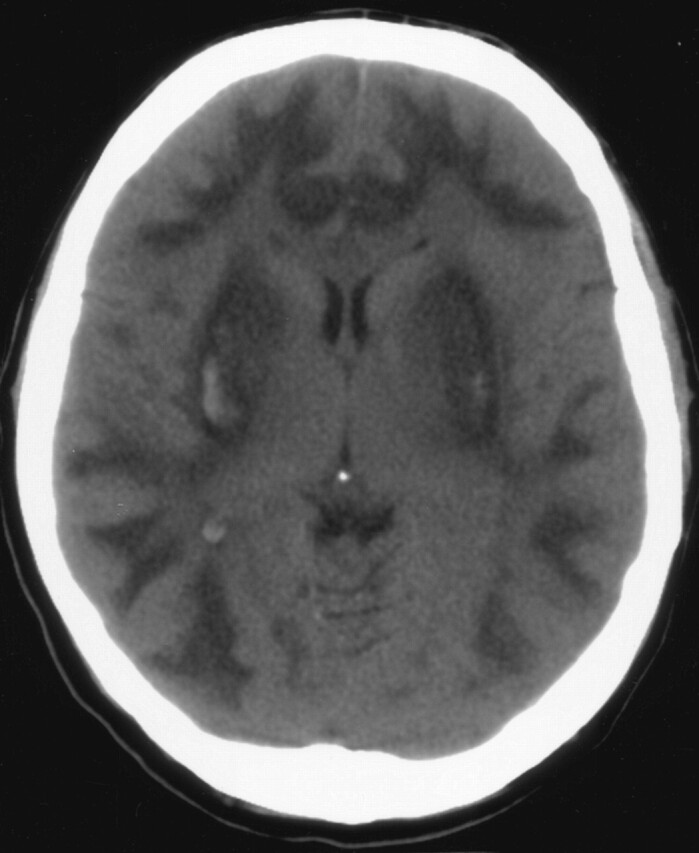
CT performed on day 5 shows low attenuation areas in subcortical white matter and both putamina with high attenuation putaminal foci consistent with hemorrhage.
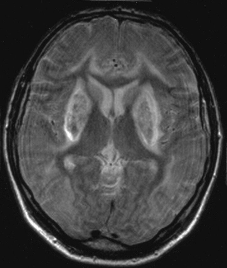
Axial fast spin-echo T2-weighted MR on day 7 shows subcortical white matter and basal ganglia hyperintensity and low-signal-intensity bilateral putaminal foci.
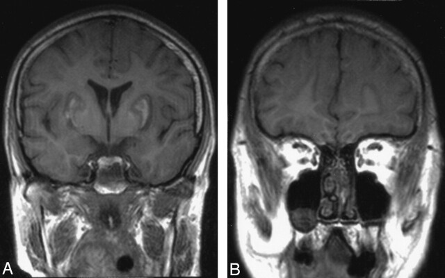
Unenhanced coronal T1-weighted image shows low signal intensity subcortical white matter and basal ganglia lesions with high-signal-intensity foci in both putamina.
Contrast-enhanced coronal T1-weighted image shows intense linear peripheral enhancement in subcortical and putaminal lesions
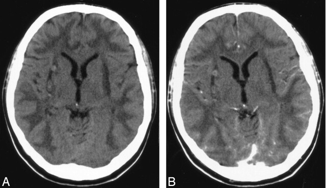
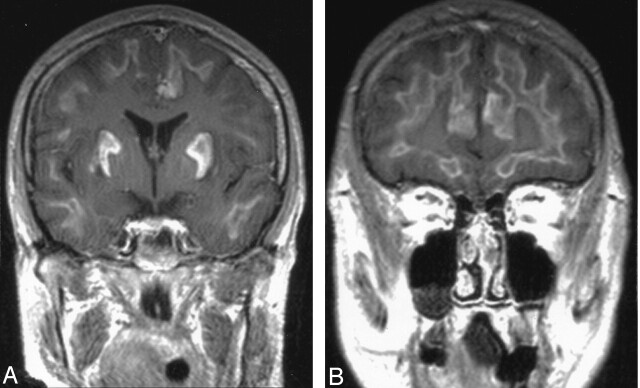
Pre- (A) and postcontrast (B) follow-up CT performed on day 24 shows bilateral putaminal volume loss and very slight enhancement of subcortical and putaminal lesions.
References
-
- Kuteifan K, Oesterle H, Tajahmady T, et al. Necrosis and haemorrhage of the putamen in methanol poisoning shown on MRI. Neuroradiology 1998;40:158–60 - PubMed
-
- Halavaara J, Valanne L, Setala K. Neuroimaging supports the clinical diagnosis of methanol poisoning. Neuroradiology 2002;44:924–28 - PubMed
-
- Hsu HH, Chen CY, Chen FH, et al. Optic atrophy and cerebral infarcts caused by methanol intoxication: MRI. Neuroradiology 1997;39:192–94 - PubMed
Publication types
MeSH terms
Substances
LinkOut - more resources
Full Text Sources
Medical