Comparative Study
. 2006 Jun 15;107(12):4834-40.
doi: 10.1182/blood-2005-08-3076. Epub 2006 Feb 16.

Phosphatidylinositol-3-phosphate kinase pathway activation protects leukemic large granular lymphocytes from undergoing homeostatic apoptosis

Affiliations
Comparative Study

Phosphatidylinositol-3-phosphate kinase pathway activation protects leukemic large granular lymphocytes from undergoing homeostatic apoptosis

Andrew E Schade et al. Blood. .

Abstract

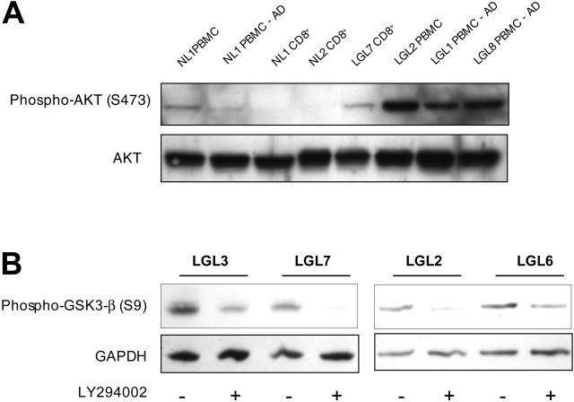
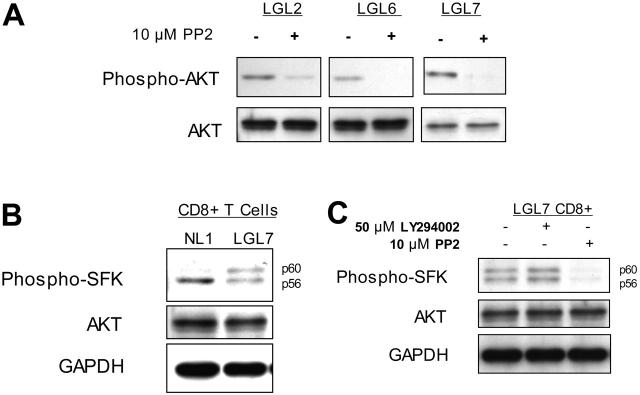
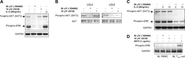
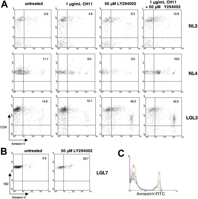
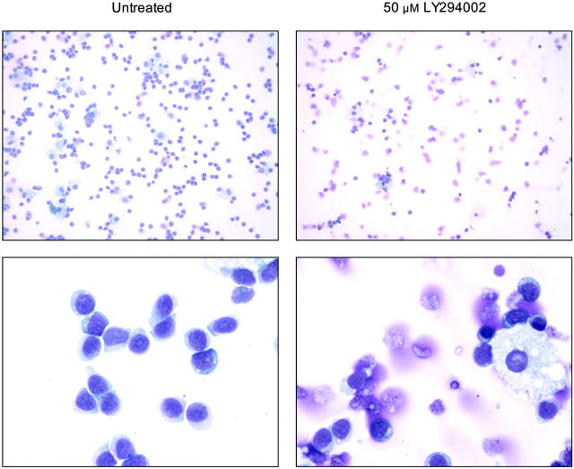
T-cell large granular lymphocytic leukemia (T-LGL) is characterized by chronic clonal lymphoproliferation of cytotoxic T lymphocytes (CTLs). Despite exhibiting phenotypic properties of antigen-activated cells, including expression of Fas and FasL, T-LGL cells accumulate and demonstrate resistance to apoptosis. We propose that increased activity of a prosurvival signaling pathway in T-LGL is responsible for attenuated apoptosis in T-LGL. Given the importance of the phosphatidylinositol-3 kinase (PI3K)-AKT pathway in regulating the balance between survival and apoptosis, we analyzed AKT activity in T-LGL cells. Compared with resting CTLs from healthy donors, patients' T-LGL cells showed higher levels of phosphorylated AKT. We demonstrate that phospho-AKT induction is dependent on the upstream activity of a Src family kinase. Since the PI3K-AKT pathway can antagonize the ability of Fas to initiate apoptosis, we hypothesized that inhibition of PI3K would lead to reacquisition of Fas sensitivity in T-LGL. Inhibition of the PI3K-AKT pathway alone led to brisk spontaneous apoptosis of T-LGL. These results suggest that T-LGL pathogenesis is dependent on activity of the PI3K-AKT pathway, without which the leukemic cells will begin to undergo spontaneous apoptosis. We propose that novel therapeutics inhibiting the PI3K-AKT axis may provide effective treatment for T-LGL.


Figures

Figure 1.
Figure 1.
PI3K-AKT pathway activity in T-LGL cells. (A) Lysates of purified CD8 T cells or PBMCs before and after adherent cell depletion, as indicated, from healthy donors and patients with T-LGL were prepared by direct addition of 2 × SDS sample buffer after a brief 15-minute incubation at 37°C and separated on a 10% SDS-PAGE gel. Following transfer to nitrocellulose membrane, samples were sequentially analyzed for phosphorylated AKT (S473) and total AKT protein via immunoblotting. Cell equivalents (3 × 10⁵) were loaded in all lanes. (B) PBMCs from 4 patients with T-LGL were incubated at 37°C for 15 minutes in the presence or absence of LY294002 (50 μM), followed by immediate addition of 2 × SDS sample buffer. Proteins were separated on an SDS-PAGE gel as in panel A, transferred to nitrocellulose, and immunoblotted for phosphorylated GSK-3β (S9). GAPDH levels were probed to indicate consistency of protein loading.
Figure 2.
Figure 2.
PI3K activity in T-LGL is dependent on the activation of a Src family kinase. (A) PBMCs from 3 patients with T-LGL were incubated at 37°C for 15 minutes in the presence or absence of the SFK inhibitor PP2 (10 μM), followed by immediate addition of 2 × SDS sample buffer. Cell equivalents (3 × 10⁵)/lane were run on a 10% SDS-PAGE gel, transferred to nitrocellulose, and immunoblotted for phosphorylated AKT (S473) and total AKT. (B) CD8+ T cells isolated from a healthy donor and a patient with T-LGL via RosetteSep were resuspended in RPMI at 5 × 10⁷ cells/mL and incubated at 37°C for 15 minutes, followed by immediate addition of 2 × SDS sample buffer and immunoblotted with an antibody specific for active SFKs. (C) CD8+ T cells isolated from a patient with T-LGL via RosetteSep were resuspended in RPMI at 5 × 10⁷ cells/mL and incubated at 37°C for 15 minutes in the presence or absence of LY294002 (50 μM) or PP2 (10 μM), as indicated. Lysates prepared as for panel B were immunoblotted for phospho-SFK. Total AKT and GAPDH protein levels are shown as loading controls.
Figure 3.
Figure 3.
ERK signaling in T-LGL is dependent on PI3K activity. (A) T-LGL PBMCs (5 × 10⁷ cells/mL) were untreated (control) or stimulated with IL-2 (20 ng/mL), LY294002 (50 μM), or U0126 (10 μM) for 15 minutes at 37°C. Lysates were prepared by immediate addition of 2 × SDS buffer. After 10% SDS-PAGE and transfer, nitrocellulose membranes were sequentially analyzed for phospho-AKT (S473) and phospho-ERK. (B) T-LGL PBMCs (5 × 10⁷ cells/mL) were untreated or stimulated with IL-2 (20 ng/mL) in the presence or absence of LY294002 (50 μM) for the indicated times at 37°C. Lysates were prepared by immediate addition of 2 × SDS buffer. After 10% SDS-PAGE and transfer, nitrocellulose membranes were sequentially analyzed for phospho-AKT (S473) and phospho-ERK. (C) T-LGL PBMCs (5 × 10⁷ cells/mL) were untreated or with LY294002 (50 μM) or U0126 (10 μM) for 15 minutes at 37°C. Lysates were prepared by immediate addition of 2 × SDS buffer. Samples were subjected to SDS-PAGE and Western blotting for phospho-AKT (S473). For all immunoblots, GAPDH or total AKT were probed to indicate consistency of protein loading between lanes on a given gel. (D) Normal PBMCs and IL-2–activated T cells (TAC) were incubated with LY294002 (50 μM) or U0126 (10 μM) for 15 minutes at 37°C, or with 1 μg/mL OKT3 for 1 minute prior to lysis in 2 × SDS sample buffer. Proteins were separated on an SDS-PAGE gel as in the previous panels, transferred to nitrocellulose, and immunoblotted for phosphorylated ERK.
Figure 4.
Figure 4.
Apoptosis induction after PI3K pathway inhibition in T-LGL. (A) Healthy-donor PBMCs and T-LGL PBMCs were incubated overnight in RPMI with 10% FCS at 37°C either untreated or treated with CH11 (anti-Fas; 1 μg/mL), LY294002 (50 μM), or CH11 (1 μg/mL) + LY294002 (50 μM), as indicated. After 20 hours the cells were stained for FITC–annexin V and PC5-CD8 and analyzed via FACS. For analysis, cells were gated on the lymphocyte population based on forward and scatter profile. The percentage of CD8+ cells that are annexin V positive are indicated by the numbers in the top right quadrants. (B) PBMCs from a T-LGL patient with a large Vβ2 clonal expansion (90% of all PBMCs) were incubated overnight in RPMI with 10% FCS at 37°C either untreated, or with 50 μM LY294002. After 20 hours, the cells were stained for FITC–annexin V and PE-Vβ2 and analyzed via FACS. For analysis, cells were gated on the Vβ2 population. The percentage of Vβ2+ cells that are annexin V positive are indicated by the numbers in the top right quadrants. (C) T-LGL PBMCs were incubated overnight in RPMI with 10% FCS at 37°C either untreated (red) or with 6.25 μM LY294002 (gray), 12.5 μM LY294002 (orange), 25 μM LY294002 (green), or 50 μM LY294002 (blue). After 16 hours the cells were stained for FITC–annexin V and PC5-CD8 and analyzed via FACS. For analysis, cells were gated on the lymphocyte population based on forward and scatter profile.
Figure 5.
Figure 5.
PI3K inhibition induces morphologic changes in T-LGL consistent with apoptosis. Untreated (control) T-LGL are noted in the left panels intermixed with occasional macrophages. The right panels show images of Wright-stained T-LGL PBMCs that had been treated for 20 hours with LY294002 (50 μM). Apoptotic cells are seen in the LY-treated T-LGL. Original magnifications: low power, × 200; high power, × 1000 (oil).

References

    1. Melenhorst JJ, Sorbara L, Kirby M, Hensel NF, Barrett AJ. Large granular lymphocyte leukaemia is characterized by a clonal T-cell receptor rearrangement in both memory and effector CD8(+) lymphocyte populations. Br J Haematol. 2001;112: 189-194. - PubMed
    1. Lamy T, Liu JH, Landowski TH, Dalton WS, Loughran TP Jr. Dysregulation of CD95/CD95 ligand-apoptotic pathway in CD3(+) large granular lymphocyte leukemia. Blood. 1998;92: 4771-4777. - PubMed
    1. Kothapalli R, Nyland SB, Kusmartseva I, Bailey RD, McKeown TM, Loughran TP Jr. Constitutive production of proinflammatory cytokines RAN-TES, MIP-1beta and IL-18 characterizes LGL leukemia. Int J Oncol. 2005;26: 529-535. - PubMed
    1. Liu JH, Wei S, Lamy T, et al. Chronic neutropenia mediated by fas ligand. Blood. 2000;95: 3219-3222. - PubMed
    1. Shvidel L, Duksin C, Tzimanis A, et al. Cytokine release by activated T-cells in large granular lymphocytic leukemia associated with autoimmune disorders. Hematol J. 2002;3: 32-37. - PubMed

Publication types

MeSH terms