Comparative Study
. 2006 Feb;2(2):e10.
doi: 10.1371/journal.ppat.0020010. Epub 2006 Feb 17.

Why do HIV-1 and HIV-2 use different pathways to develop AZT resistance?

Affiliations
Comparative Study

Why do HIV-1 and HIV-2 use different pathways to develop AZT resistance?

Paul L Boyer et al. PLoS Pathog. 2006 Feb.

Abstract

The human immunodeficiency virus type 1 (HIV-1) develops resistance to all available drugs, including the nucleoside analog reverse transcriptase inhibitors (NRTIs) such as AZT. ATP-mediated excision underlies the most common form of HIV-1 resistance to AZT. However, clinical data suggest that when HIV-2 is challenged with AZT, it usually accumulates resistance mutations that cause AZT resistance by reduced incorporation of AZTTP rather than selective excision of AZTMP. We compared the properties of HIV-1 and HIV-2 reverse transcriptase (RT) in vitro. Although both RTs have similar levels of polymerase activity, HIV-1 RT more readily incorporates, and is more susceptible to, inhibition by AZTTP than is HIV-2 RT. Differences in the region around the polymerase active site could explain why HIV-2 RT incorporates AZTTP less efficiently than HIV-1 RT. HIV-1 RT is markedly more efficient at carrying out the excision reaction with ATP as the pyrophosphate donor than is HIV-2 RT. This suggests that HIV-1 RT has a better nascent ATP binding site than HIV-2 RT, making it easier for HIV-1 RT to develop a more effective ATP binding site by mutation. A comparison of HIV-1 and HIV-2 RT shows that there are numerous differences in the putative ATP binding sites that could explain why HIV-1 RT binds ATP more effectively. HIV-1 RT incorporates AZTTP more efficiently than does HIV-2 RT. However, HIV-1 RT is more efficient at ATP-mediated excision of AZTMP than is HIV-2 RT. Mutations in HIV-1 RT conferring AZT resistance tend to increase the efficiency of the ATP-mediated excision pathway, while mutations in HIV-2 RT conferring AZT resistance tend to increase the level of AZTTP exclusion from the polymerase active site. Thus, each RT usually chooses the pathway best suited to extend the properties of the respective wild-type enzymes.


Conflict of interest statement

Competing interests. The authors have declared that no competing interests exist.

Figures

Figure 1
Figure 1. Processivity of Wild-Type HIV-1 and HIV-2 RTs
As described in Materials and Methods, a 5′ end-labeled primer was annealed to single-strand M13mp18 DNA, then extended with wild-type HIV-1 RT or HIV-2 RT in the presence of 10.0 μM of each dNTP and unlabeled poly(rC)•oligo(dG), which acts as a “cold trap.” The cold trap limits extension to one round of polymerization. The location of the size marker bands (in nucleotides) are shown on the left.
Figure 2
Figure 2. Inhibition of RT Polymerization by AZTTP
The assay was done as previously described [1]. Normal dNTPs were present in the reaction at 10.0 μM each. Increasing concentrations of AZTTP were added, and the level of radioactive [α−32P]dCTP incorporated into the template/primer was measured. The level of radioactivity incorporated in the absence of analog was considered 100% activity; the other reactions were normalized to this value. The error bars are included; however, they are partially obscured by the data symbols. RT1 and RT2 designate the HIV RT backbone in which the Q151M mutation was made: HIV-1 or HIV-2.
Figure 3
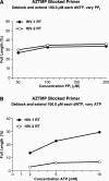
Figure 3. Excision/Extension of AZTMP by HIV-1 RT and HIV-2 RT
As described in Materials and Methods, a 5′ end-labeled primer was annealed to a template. The 3′ end of the primer was blocked by the addition of AZTMP. (A) The excision/extension assays were performed with AZTMP-blocked primers in the presence of high levels of dNTPs (100.0 μM each), which favors formation of the ternary complex (RT/DNA/dNTP) and the indicated concentrations of NaPPi (50.0, 100.0, 200.0 μM). All experiments were done at least in duplicate; a typical result is shown. (B) Excision/extension of an AZTMP-blocked primer using high levels of dNTPs (100.0 μM each dNTP) and the indicated concentrations of ATP (2.0, 5.0, 10.0 mM). All experiments were done at least in duplicate; a typical result is shown.
Figure 4
Figure 4. Comparison of the First 240 Amino Acids of HIV-1 RT (Strain BH10) and HIV-2 RT (Strain ROD)
The triad of aspartic acid residues critical for polymerase activity is shown in red. The amino acid residues that are altered as part of the Q151M complex in HIV-1 RT are shown in blue; residues 214, 215, and 219 are shown in plum.
Figure 5
Figure 5. Polymerase Active Site Regions of HIV-1 RT and of HIV-2 RT
(A) The HIV-1 RT complex with an AZTMP-terminated primer trapped at the N site (PDB code 1N6Q). (B) Nucleic acid from (A) superposed on the structure of unliganded HIV-2 RT (PBD code 1MU2). The side chains of amino acid residues proximal to the azido group of AZTMP are shown as white spheres, which represent the Van der Waals volumes. The side chains of amino acid residues at the interface of several secondary structure elements of RT are shown in different colors: 209–215 of the large subunit (green spheres), 41–46 of the large subunit (magenta spheres), 116–117 of the large subunit (red spheres), and the N-terminus of the large subunit (cyan spheres). The proposed ATP-binding regions are shown as semitransparent yellow ellipsoids. The direction of the nucleophilic attack of ATP on the AZTMP-terminated primer is indicated by a black arrow.
Figure 6
Figure 6. A Comparison of the Structure of an HIV-1 RT/DNA Complex with the Structure of Unliganded HIV-2 RT
In the HIV-1 RT complex (PDB code 1N6Q), shown in yellow, the 3′ end of the primer is in the N site, unliganded HIV-2 RT (PDB code 1MU2) is shown in magenta. The residues used for the superposition were 107′112 and 155′215. The Van der Waal radii for the atoms in the amino acid residues at positions 117 and 214 are shown as spheres: yellow for HIV-1 RT and blue for HIV-2 RT. The superposition highlights two major differences between the two enzymes: a) the interaction between the N terminus and residue Ser117 of p66 in HIV-1 RT (red dotted line) is absent in HIV-2 RT because in HIV-2 RT the N terminus has moved away from residue 117 (cyan dotted line); b) in HIV-1 RT, Ser117 interacts with Leu214 1 RT in a way that differs from the interaction of Ser117 and the bulkier Phe214 in HIV-2 RT.
Figure 7
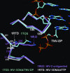
Figure 7. Position of the Side Chain of Amino Acid Residue K219 in HIV-1 RT and K220 in HIV-2 RT
Unliganded HIV-2 RT (shown in magenta; PDB code 1MU2) and HIV-1 RT/DNA/dTTP (shown in white with the dTTP and DNA omitted for clarity; PDB code 1RTD), is superimposed on the Cα protein backbone of HIV-1 RT/DNA/tenofovir-diphosphate (shown in cyan; PDB code 1TO5). The residues used for the superposition were 107–112 and 155–215. The alignment shows that K220 of HIV-2 RT is the residue structurally equivalent to K219 in HIV-1 RT.
Figure 8
Figure 8. Superposition of HIV-1 RT and HIV-2 RT Showing the Possible Effects of a Phe116Tyr Mutation
Unliganded HIV-2 RT (PDB code 1MU2) is shown in yellow, unliganded HIV-1RT (PDB code 1DLO) is shown in cyan, and HIV-1 RT/DNA/dTTP (PDB code 1RTD) is shown in red. The superposition shows that a Phe116Tyr mutation in HIV-1 RT would affect the interactions of position 116 with the main-chain carbonyl of Lys73 in the fingers subdomain of HIV-1 RT (dotted line). However, the same mutation in HIV-2 RT is not likely to cause similar interactions of Tyr116 with the fingers subdomain of HIV-2 because the differences in the position of the small helix that carries residues 115–117 in HIV-2 RT make the interaction unlikely.

References

    1. Sarafianos SG, Das K, Hughes SH, Arnold E. Taking aim at a moving target: Designing drugs to inhibit drug-resistant HIV-1 reverse transcriptases. Curr Opin Struc Biol. 2004;14:716–730. - PubMed
    1. Arion D, Kaushik N, McCormick S, Borkow G, Parniak MA. Phenotypic mechanism of HIV-1 resistance to 3′-azido-3′-deoxythymidine (AZT): Increased polymerization processivity and enhanced sensitivity to pyrophosphate of the mutant viral reverse transcriptase. Biochemistry. 1998;37:15908–15917. - PubMed
    1. Boyer PL, Sarafianos SG, Arnold E, Hughes SH. Selective excision of AZTMP by drug-resistant human immunodeficiency virus reverse transcriptase. J Virol. 2001;75:4832–4842. - PMC - PubMed
    1. Meyer PR, Matsuura SE, So AG, Scott WA. Unblocking of chain-terminated primer by HIV-1 reverse transcriptase through a nucleotide-dependent mechanism. Proc Natl Acad Sci U S A. 1998;95:13471–13476. - PMC - PubMed
    1. Boyer PL, Sarafianos SG, Arnold E, Hughes SH. Nucleoside analog resistance caused by insertions in the fingers of human immunodeficiency virus type 1 reverse transcriptase involves ATP-mediated excision. J Virol. 2002;76:9143–9151. - PMC - PubMed

Publication types