

Echinococcosis in Tibetan populations, western Sichuan Province, China
- PMID: 16485472
- PMCID: PMC3367622
- DOI: 10.3201/eid1112.050079
Echinococcosis in Tibetan populations, western Sichuan Province, China
Abstract
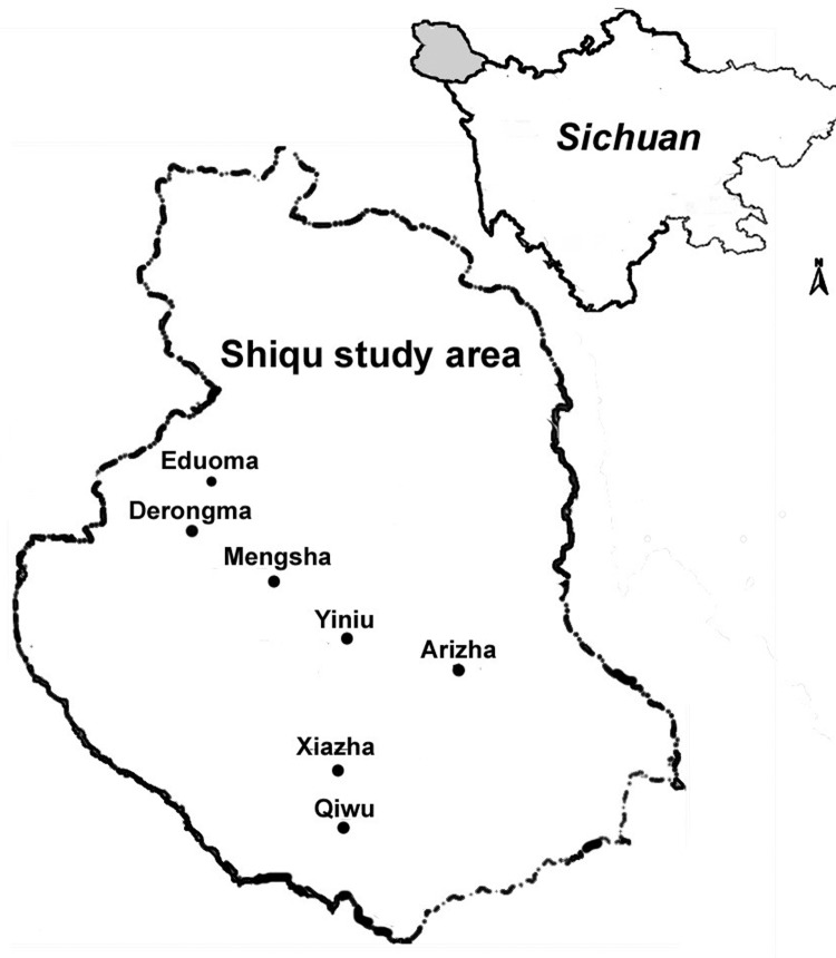
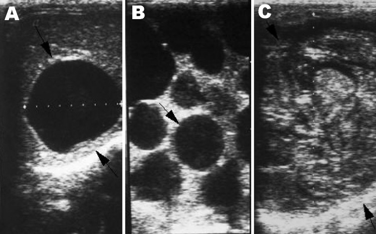
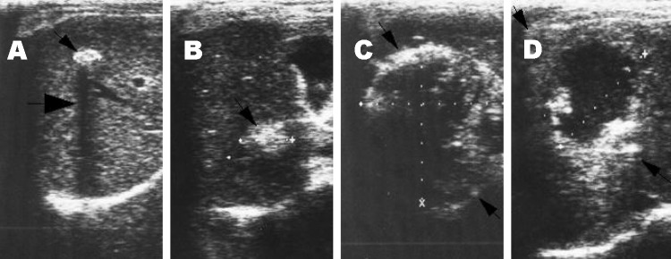
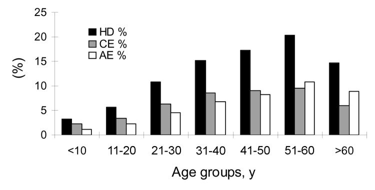
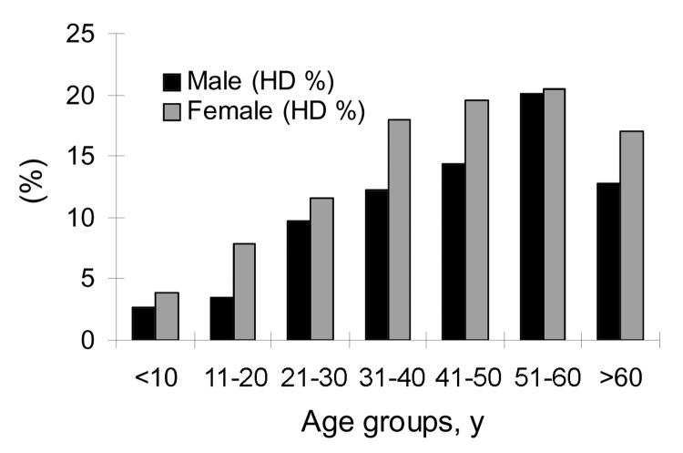
We screened 3,199 people from Shiqu County, Sichuan Province, China, for abdominal echinococcosis (hydatid disease) by portable ultrasound combined with specific serodiagnostic tests. Both cystic echinococcosis (CE) (Echinococcus granulosus infection) and alveolar echinococcosis (AE) (E. multilocularis) were co-endemic in this area at the highest village prevalence values recorded anywhere in the world: 12.9% were infected with one or the other form (6.8% CE and 6.2% AE). Prevalences of both CE and AE were significantly higher in female than male patients and increased with the age of the person screened. Pastoral herdsmen were at highest risk for infection (prevalence 19.0%). Prevalence of CE varied in 5 townships from 0% to 12.1%, whereas AE prevalence ranged from 0% to 14.3%. Risk factors associated with both infections included the number of owned dogs, frequency of contact with dogs, and sources of drinking water.
Figures
References
-
- Schantz PM, Chai JJ, Craig PS, Echert J, Jenkins DJ, Macpherson CNL, et al. Epidemiology and control of hydatid disease. In: Thompson RCA, Lymbery AJ, editors. Echinococcosis and hydatid disease. Wallingford, UK: CAB International; 1995. p. 233–331.
-
- Jiang C. Alveolar echinococcosis in China. Chin Med J (Engl). 1998;111:470–5. - PubMed
-
- Qiu JM, Liu FJ, Schantz PM, Ito A, Delker C, He JG, et al. Epidemiological study on human hydatidosis in Tibetan region of Western Sichuan. Chin J Zoonoses. 2000;10:77–80.
-
- The committee of compiling Shiqu County annals. The Shiqu annals. Chengdu, Sichuan Province, China: Sichuan People's Press; 2000. p. 1–3.
Publication types
MeSH terms
Grants and funding
LinkOut - more resources
Full Text Sources