. 2006 Mar;143(3):445-51.
doi: 10.1111/j.1365-2249.2006.03020.x.

The local immune response in ulcerative lesions of Buruli disease

Affiliations

The local immune response in ulcerative lesions of Buruli disease

A E Kiszewski et al. Clin Exp Immunol. 2006 Mar.

Abstract

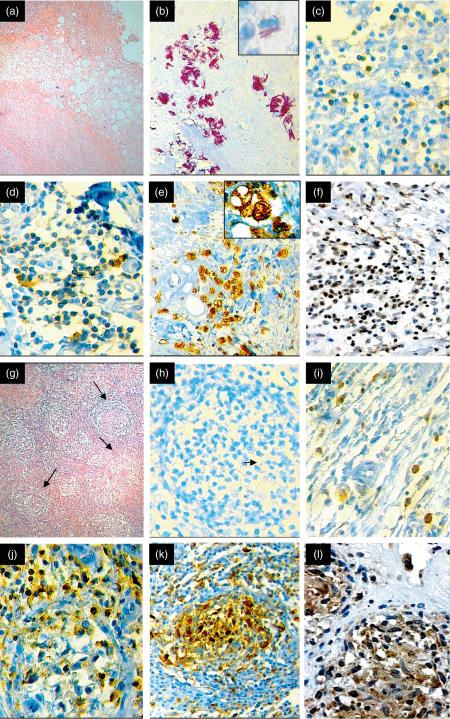
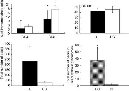
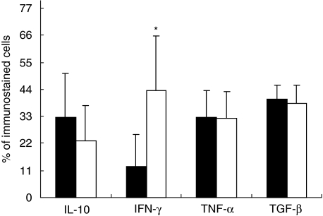
Buruli disease (BU) is a progressive necrotic and ulcerative disease of the skin and subcutaneous tissue caused by Mycobacterium ulcerans. BU is considered the third most common mycobacterial disease after tuberculosis and leprosy. Three clinical stages of the cutaneous lesions have been described in BU: pre-ulcerative, ulcerative and healed lesions. In this study we used immunohistochemistry and automated morphometry to determine the percentage of macrophages and of CD4/CD8 lymphocytes and their expression of interferon (IFN)-gamma, interleukin (IL)-10, tumour necrosis factor (TNF)-alpha and transforming growth factor (TGF)-beta. Expression of these cytokines was correlated with the inflammatory response evaluated by histopathology. All the studied BU ulcerative cases showed extensive necrosis and chronic inflammation. The most important feature was the presence or absence of granulomas co-existing with a mixed pro-inflammatory/anti-inflammatory cytokine balance. When granulomas were present significantly higher expression of IFN-gamma was seen, whereas in ulcerative lesions without granulomas there was increased expression of IL-10 and significantly higher bacillary counts. These features correlated with the chronicity of the lesions; longer-lasting lesions showed granulomas. Thus, granulomas were absent from relatively early ulcerative lesions, which contained more bacilli and little IFN-gamma, suggesting that at this stage of the disease strong suppression of the protective cellular immune response facilitates proliferation of bacilli.


Figures

Fig. 1
Fig. 1
Representative histopathological and immunohistochemical features in Buruli disease ulcers (BU). (a) Low-power photomicrograph (× 40) from a representative ulcerative BU lesion without granulomas. Extensive coagulative necrosis in the subcutaneous tissue involving the adipose and muscular tissue. (b) This lesion has a massive number of extracellular and occasional intracellular acid-fast bacilli demonstrated by Ziehl–Neelsen staining (× 400). In the insert, intracellular bacilli are shown in a vacuolated macrophage (× 1000). (c) This kind of lesion has relatively few CD4 T lymphocytes (× 200). (d) More CD8 T lymphocytes are present in this ulcerative lesion without granulomas (× 200). (e) Numerous CD68-positive macrophages are present in this lesion (× 100). In the insert, strong positivity in a vacuolated macrophage (× 1000). (f) Some IFN-γ-positive cells are seen in this BU ulcerative lesion without granulomas (× 100). (g) Low power photomicrograph (× 40) from a representative BU ulcerative case with well-formed granulomas (arrows). (h) This granulomatous lesion shows scarce acid-fast bacilli (arrow) (× 200). (i) Like non-granulomatous ulcerative lesions, granulomatous lesions also show fewer CD4 cells, with numerous CD8 cells (j) and numerous macrophages (k). (i) These granulomas show more diffusely distributed IFN-γ immunostained cells than are seen in pure ulcerative lesions (compare with f) (× 400).
Fig. 2
Fig. 2
Right and left upper panel show the percentages of CD4 cells, CD8 cells, and macrophages (CD68) determined by immunohistochemistry and automated morphometry in the subcutaneous inflammatory infiltrate from non-granulomatous ulcerative lesions (black bars) or ulcers with granulomas (white bars). Five randomly chosen fields at 200× magnification were studied in each of the 11 cases of Buruli disease. Left lower panel shows the number of acid fast bacilli in non-granulomatous ulcerative lesions (U, black bars) or ulcers with granulomas (UG, white bars), while the right lower panel shows the numbers of extracellular (EC) and intracellular (IC) bacilli in macrophages from ulcers without granulomatous lesions. Results are expressed as the mean and standard deviation. The asterisk means statistical significance.
Fig. 3
Fig. 3
Percentages of cells immunostained for interleukin (IL)-10, interferon (IFN)-γ, tumour necrosis factor (TNF)-α and transforming growth factor (TGF)-β in the inflammatory infiltrate located in the subcutaneous tissue of ulcerative lesions without granulomas (black bars) or ulcers with granulomatous lesions (white bars). Five randomly chosen fields at 200× magnification were studied through automated image analysis in each of the 11 cases of Buruli disease. Results are expressed as the mean and standard deviation. The asterisk means statistical significance.

References

    1. Asiedu K, Sherpbier R, Raviglione MC. WHO Global Buruli Ulcer Initiative Report. Geneva, Switzerland: World Health Organization; 2000. Boruli ulcer Mycobacterium ulcerans infection.
    1. van der Werf TS, van der Graff WTA, Tappero JW, Asiedu K. Mycobacterium ulcerans infection. Lancet. 1999;354:1013–18. - PubMed
    1. Meyers WM, Shelly WM, Connor DH, Meyers EK. Human Mycobacterium ulcerans infections developing at sites of trauma to skin. Am J Trop Med Hyg. 1974;23:919–23. - PubMed
    1. Marsollier I, Robert R, Aubry J, et al. Acquatic insects as a vector for Mycobacterium ulcerans. Appl Environ Microbiol. 2002;68:4623–8. - PMC - PubMed
    1. Marsollier L, Aubry J, Coutanceau E, et al. Colonization of the salivary glands of Naucoris cimicoides by Mycobacterium ulcerans requires host plasmatocytes and a macrolide toxin, mycolactone. Cell Microbiol. 2005;7:935–43. - PubMed

Publication types

MeSH terms