. 2006 May 1;30(4):1401-13.
doi: 10.1016/j.neuroimage.2005.10.053. Epub 2006 Feb 17.

Processing stages underlying word recognition in the anteroventral temporal lobe

Affiliations

Processing stages underlying word recognition in the anteroventral temporal lobe

Eric Halgren et al. Neuroimage. .

Abstract

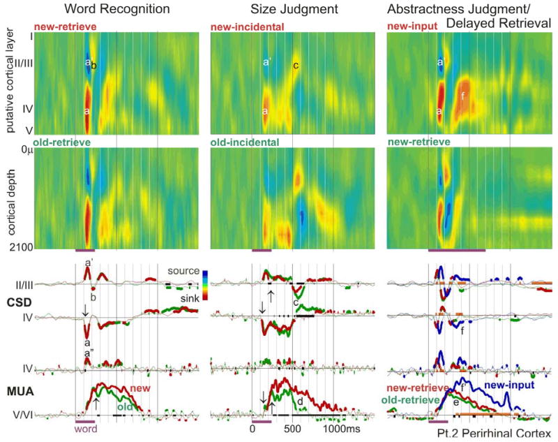
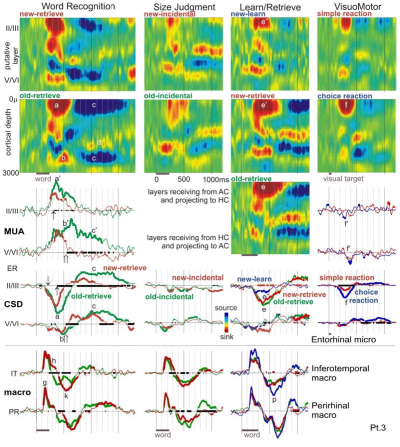
The anteroventral temporal lobe integrates visual, lexical, semantic and mnestic aspects of word processing, through its reciprocal connections with the ventral visual stream, language areas, and the hippocampal formation. We used linear microelectrode arrays to probe population synaptic currents and neuronal firing in different cortical layers of the anteroventral temporal lobe, during semantic judgments with implicit priming and overt word recognition. Since different extrinsic and associative inputs preferentially target different cortical layers, this method can help reveal the sequence and nature of local processing stages at a higher resolution than was previously possible. The initial response in inferotemporal and perirhinal cortices is a brief current sink beginning at approximately 120 ms and peaking at approximately 170 ms. Localization of this initial sink to middle layers suggests that it represents feedforward input from lower visual areas, and simultaneously increased firing implies that it represents excitatory synaptic currents. Until approximately 800 ms, the main focus of transmembrane current sinks alternates between middle and superficial layers, with the superficial focus becoming increasingly dominant after approximately 550 ms. Since superficial layers are the target of local and feedback associative inputs, this suggests an alternation in predominant synaptic input between feedforward and feedback modes. Word repetition does not affect the initial perirhinal and inferotemporal middle layer sink but does decrease later activity. Entorhinal activity begins later (approximately 200 ms), with greater apparent excitatory post-synaptic currents and multiunit activity in neocortically projecting than hippocampal-projecting layers. In contrast to perirhinal and entorhinal responses, entorhinal responses are larger to repeated words during memory retrieval. These results identify a sequence of physiological activation, beginning with a sharp activation from lower level visual areas carrying specific information to middle layers. This is followed by feedback and associative interactions involving upper cortical layers, which are abbreviated to repeated words. Following bottom-up and associative stages, top-down recollective processes may be driven by entorhinal cortex. Word processing involves a systematic sequence of fast feedforward information transfer from visual areas to anteroventral temporal cortex followed by prolonged interactions of this feedforward information with local associations and feedback mnestic information from the medial temporal lobe.


Figures

Figure 1
Figure 1. Locations of recording sites in MRIs taken with the electrodes in situ
Laminar probes are indicated by oblique arrows, and macroelectrode contacts by vertical arrows. The white MRI artifacts artifacts lateral to the probes are due to the clinical contacts (larger than the actual electrodes). Patient 1, inferotemporal cortex (IT): laminar tip in the lateral aspect of the right fusiform g., medial bank of lateral occipito-temporal s. (coordinates 38 lateral, 22 posterior, 11 down) (Talairach and Tournoux, 1988). Patient 2, perirhinal cortex (PR): laminar tip in the lateral aspect of the right parahippocampal g., medial bank of collateral s. (coordinates 31, -22, -16). Patient 3, entorhinal cortex (ER): laminar tip in the medial aspect of the left parahippocampal g. (coordinates -23, -17, -25). The left macro-electrode (↓) is in the crown of the inferior temporal gyrus (probable IT) and the right macrorecording (↑) is in the fundus of the collateral sulcus, in or near PR. Some MRIs are displayed with inverted contrast to maximize electrode visibility.
Figure 2
Figure 2. Inferotemporal population synaptic activity evoked by words in memory tasks
Responses to Word Recognition, Size Judgment and Verb Conjugation are arranged in columns. Averaged CSD color maps (upper two rows) show an initial transmembrane current sink in putative layer IV (red area, a), with a return source in more superficial layers (blue area, a’). These sinks and sources are longer duration to new words (b, b’). The sink may invert to a source, and vice versa, several times from ~300–800ms (c, c’), culminating in a sustained superficial sink (d) and middle layer source (d’). The same phenomena can be seen in the averaged CSD waveforms from selected superficial and middle layers in the lower 3 rows. Purple bars below x-axes indicate word presentation periods. The x-axis is thickened when new and old words evoke significantly different activity; CSD waveforms are thickened when significantly different from zero (2-tailed p<0.01). The synaptic response begins at ~120ms (↓), but differential activity to word repetition does not begin until ~220ms after stimulus onset (↑).
Figure 3
Figure 3. Perirhinal activity during memory tasks
Words evoke an initial sharp middle layer transmembrane current sink (a) and superficial source (a’), seen in the CSD color maps (upper two rows) and waveforms (middle two rows). The sink likely represents a population EPSC, since it is associated with increased population firing (lower two rows), especially in the middle layer (a”). This is followed by a sink in superficial layers (b), and then variable activity including a very late superficial sink during Size Judgments (c). Population firing and synaptic activity begins at ~120ms (↓), but differential activity to word repetition does not begin until ~220ms after stimulus onset (↑) when new words evoke stronger sustained firing in deep layers until 800–1100ms (d). Similar patterns are observed regardless of whether word repetition is explicit (Word Recognition, first column), or implicit (Size Judgment, second column). The third column also shows an explicit word recognition task, but with a single word presentation during Abstractness Judgments, and an ~1hr delay before Delayed Retrieval. In Word Recognition and Size Judgment, old words are seen several times with delays of ~1min. Although the repetition effect is smaller and later in Delayed Retrieval, it is still present (e). Note that compared to new words during the output period, new words during the input task evoke a larger middle layer sink (f) and more population firing (f’). The x-axis is thickened when responses to new and old words are significantly different from each other; CSD and MUA waveforms are thickened when they are significantly different from zero (2-tailed p<0.01). The thick orange baseline indicates significant differences between responses evoked by new words at input versus retrieval.
Figure 4
Figure 4. Entorhinal population synaptic activity and neuronal firing
CSD color maps (upper rows) show the spatiotemporal patterns of population synaptic activity across four tasks (arrayed in columns)., Waveforms below CSD maps show simultaneous MUA, CSD and macro-electrode recordings (third through fifth rows). The most prominent early activity during explicit Word Recognition is a superficial sink starting at ~200ms (↓) and peaking at ~370ms (a) with strongly increased firing in superficial and deep layers (a’). Sinks in deeper layers (b) with accompanying MUA (b’) also occur during this time. Both deep and superficial sinks are followed by sources in the same layers (c), beginning at ~700ms after the stimulus and continuing for ~900ms. The deep source is surrounded by sinks (e.g., d). Population firing in deep layers continues at a high level during this period (c’), whereas that in superficial layers returns to baseline. All components of the response are larger to old as compared to new words, with the CSD and MUA responses diverging in superficial layers at ~300–400ms (↑), and in deep layers at ~500–600ms (⇑). A broadly similar but substantially weaker spatiotemporal pattern is seen to implicit word repetition during Size Judgments (second column). In Learn/Retrieve (third column), subjects were explicitly asked to memorize the words during Learn, and then were presented with forced-choice explicit recognition. In addition to showing a larger synaptic response to old (e) as compared to new (e’) words during Retrieval, the response is larger to new words during Retrieval (e’) as opposed to new words during Learn (e”). In the fourth column, choice VisuoMotor reactions evoke a similar superficial sink (f) as in Word Recognition, but with less than half the amplitude or duration (a). The difference in neuronal firing is more striking, with VisuoMotor reactions evoking a decrease in firing (f’) whereas Word Recognition evokes a sustained increase (a’, b’, c’). Bipolar potential recordings from macro-electrodes in left IT and right PRshow distinct spatiotemporal patterns that resemble the IT and PR laminar recordings, beginning with a sharp initial peak before 200ms (g), followed by two components from ~240 to 620ms (h, k) that distinguish new and old words. In contrast to the ER laminar recordings, IT and PR potential gradients are largest during Learning (p). The x-axis is thickened when two conditions evoke significantly different activity (black lines for new versus old words, or choice versus simple reactions; orange lines for new-learn vs. new-retrieve in Learn/Retrieve); CSD and MUA waveforms are thickened when each is significantly different from zero (2-tailed p<0.01). MUA responses are only shown for Word Recognition and VisuoMotor tasks, because they were given on the first day of testing when the MUA recordings were higher quality. Macro-electrode recordings were not obtained for the VisuoMotor task.

Similar articles

Cited by

References

    1. Allison T, Puce A, Spencer DD, McCarthy G. Electrophysiological studies of human face perception. I: Potentials generated in occipitotemporal cortex by face and non-face stimuli. Cereb Cortex. 1999;9:415–430. - PubMed
    1. Bancaud J, Brunet-Bourgin F, Chauvel P, Halgren E. Anatomical origin of déjà vu and vivid 'memories' in human temporal lobe epilepsy. Brain. 1994;117:71–90. - PubMed
    1. Barbas H, Rempel-Clower N. Cortical structure predicts the pattern of corticocortical connections. Cereb Cortex. 1997;7:635–646. - PubMed
    1. Barth DS, Di S. Three-dimensional analysis of auditory-evoked potentials in rat neocortex. J Neurophysiol. 1990;64:1527–1536. - PubMed
    1. Bartolomei F, Barbeau E, Gavaret M, Guye M, McGonigal A, Regis J, Chauvel P. Cortical stimulation study of the role of rhinal cortex in deja vu and reminiscence of memories. Neurology. 2004;63:858–864. - PubMed

Publication types

MeSH terms