Review
. 2006 Mar;11(3):115-23.
doi: 10.1016/j.tplants.2006.01.008. Epub 2006 Feb 20.

Exploring the ESCRTing machinery in eukaryotes

Affiliations
Review

Exploring the ESCRTing machinery in eukaryotes

Verena Winter et al. Trends Plant Sci. 2006 Mar.

Abstract

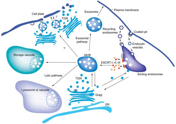
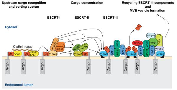
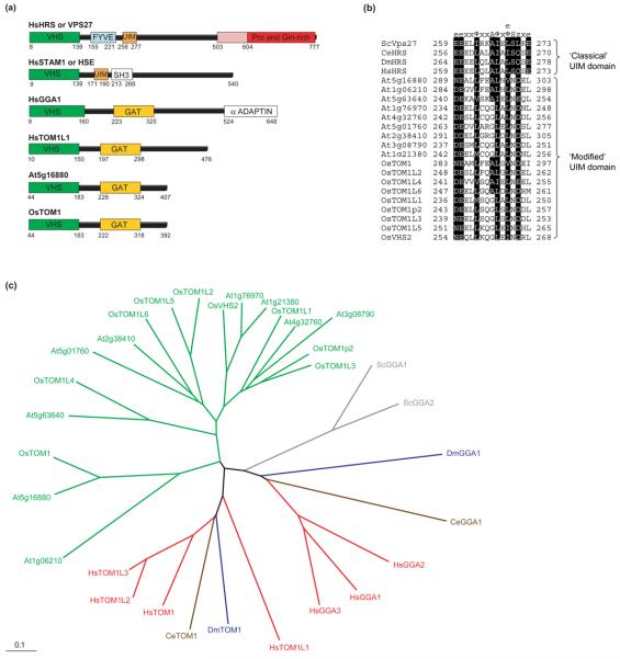
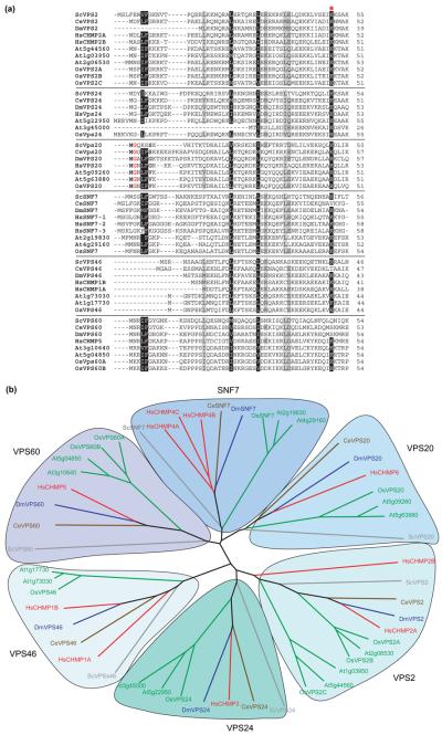
The profile of protein sorting into multivesicular bodies (MVBs) has risen recently with the identification of three heteromeric complexes known as ESCRT-I,-II,-III (Endosomal Sorting Complex Required for Transport). Genetic analyses in yeast have identified up to 15 soluble class E VPS (vacuolar protein sorting) proteins that have been assigned to the ESCRT machinery and function in cargo recognition and sorting, complex assembly, vesicle formation and dissociation. Despite their functional importance in yeast and mammalian cells, little is known about their presence and function in other organisms including plants. We have made use of the fully sequenced genomes of Arabidopsis thaliana and Oryza sativa, Drosophila melanogaster and Caenorhabditis elegans to explore the identity, structural characteristics and phylogenetic relationships of proteins assigned to the ESCRT machinery.


Figures

Figure 1
Figure 1
Characteristics and roles of multivesicular bodies (MVBs). MVBs are endocytic intermediates that enclose, in a limiting membrane, luminal vesicles that originate by invagination at sorting endosomes. MVBs function at the cross road between endocytosis, exocytosis and transport to lysosomes or vacuoles and serve as prevacuolar compartments and precursors of exosomes. Sorting into MVBs can determine the delivery of transmembrane proteins into secretory lysosomes, and thus MVBs are involved in the activity control of endocytosed receptors. Furthermore, MVBs might also serve as storage compartments and carriers for morphogens. Invagination and protein sorting to MVBs requires the coordinated action of three multiprotein complexes, ESCRT-I, -II, -III (see Figure 2).
Figure 2
Figure 2
Model of the ESCRT-dependent protein sorting and concentration machinery in the MVB vesicle formation pathway. Monoubiquitinated transmembrane proteins are recognized by the upstream cargo sorting system of endosomes via a series of ubiquitin- and clathrin-binding proteins (GGAs, VPS27 and TOM1) that recruit ESCRT-I. ESCRT-I is able to bind ubiquitin via VPS23. The formation of ESCRT-I activates ESCRT-II, which initiates the formation of the ESCRT-III complex through the interaction of VPS22, VPS25 and VPS36 (ESCRT-II) with VPS20 (ESCRT-III). Concurrent with inward membrane budding the ubiquitin tags of the cargo proteins are removed by the deubiquitinating enzyme DOA4 and the ESCRT-III complex components dissociate from the endosomal membrane through the AAA-type ATPase activity of VPS4. Experimentally proven protein–protein interactions are indicated by arrows. The model also shows the known protein–protein interactions within the ESCRT complex. Dimerizations are symbolized as double boxes. The myristoylation of VPS20 is symbolized with a black triangle.
Figure 3
Figure 3
Structural similarities and phylogenetic analysis of components of the upstream cargo recognition and sorting system. (a) Domain organization of the human HRS or VPS27, STAM1, GGA1 and TOM1L1 proteins compared with a representative VHS-GAT domain protein of Arabidopsis (At5g16880) and rice (OsTOM1). The 100 amino acids of the Pro- and Gln-rich domain of HRS, which is essential for the localization to endosomes, are depicted in pink. (b) Comparison of the UIM domain of VPS27 and HRS proteins and the equivalent domain of VHS-GAT proteins of Arabidopsis and rice. Highly conserved residues are highlighted in black. Note the deviations from the ‘classical’ UIM domain with the consensus of e-e-x-x-φ-x-x-A-φ-x-e/φ-S-z-x-e, where e stands for negatively charged, φ for hydrophobic, x for helix-favoring and z for polar or hydrophobic residues. Ala and Ser are invariant. The VHS-GAT-containing putative plant homologs have an altered UIM motif at the same position: the invariant Ala and Ser are exchanged to Gly and Asp, respectively and the last negatively charged amino acid is replaced by a hydrophobic one. (c) Neighbor-joining phylogenetic tree generated with the VHS-GAT domains showing the predicted relationship between the yeast, Drosophila, Caenorhabditis elegans and human GGA and TOM1 proteins and their homologs in plants. The phylogram is based on an alignment prepared using Clustal X 1.81 and was drawn using Tree-View 1.6.6. The color-coded nomenclature is similar to that used in Table S1 in the Supplementary material and the first two letters of each protein name represent the organism: Sc, Saccharomyces cerevisiae (gray); Dm, Drosophila melanogaster (blue); Ce, Caenorhabditis elegans (brown); At, Arabidopsis thaliana (green); Os, Oryza sativa (green); and Hs, Homo sapiens (red).
Figure 4
Figure 4
Protein alignments and phylogenetic trees of the ESCRT-III complex components. (a) Alignment of the first 50 amino acids of all the ‘classical (VPS2, SNF7, VPS24, VPS20)’ and nonclassical (VPS46, VPS60) proteins of the ESCRT-III complex. Highly conserved residues are highlighted in black. The Gly highlighted in red font and the surrounding residues of the VPS20 class highlighted in bold mark the N-terminal myristoylation site. The red asterisk marks the conserved Lys (K49), which is important for lipid and membrane interactions. (b) Neighbor-joining phylogenetic tree with 1000 bootstrap replicates generated with all the SNF7 domain-containing ESCRT-III complex components. The tree is based on a protein alignment prepared with Clustal × 1.81 and was drawn using Tree-View 1.6.6. The color-coded nomenclature is similar to that used in Table S4 in the Supplementary material. The six SNF7-domain protein subclasses are highlighted and named after their yeast protagonists. Note the expansion of members in the plant lineages and the plant-specific VPS2 subclass.

References

    1. Palade GEA. A small particulate component of the cytoplasm. J. Biophys. Biochem. Cytol. 1955;1:59–67. - PMC - PubMed
    1. Tanchak MA, Fowke LC. The morphology of multivesicular bodies in soybean protoplasts and their role in endocytosis. Protoplasma. 1987;138:173–182.
    1. Marcote MJ, et al. Membrane transport in the endocytic pathway: animal versus plant cells. Protoplasma. 2000;210:123–132.
    1. Jiang L, et al. Multivesicular bodies: a mechanism to package lytic and storage functions in one organelle? Trends Cell Biol. 2002;12:362–367. - PubMed
    1. Tse YC, et al. Identification of multivesicular bodies as prevacuolar compartments in Nicotiana tabacum BY-2 cells. Plant Cell. 2004;16:672–693. - PMC - PubMed

Publication types