. 2006 Feb 18;34(4):1102-11.
doi: 10.1093/nar/gkj512. Print 2006.

Novel thermostable Y-family polymerases: applications for the PCR amplification of damaged or ancient DNAs

Affiliations

Novel thermostable Y-family polymerases: applications for the PCR amplification of damaged or ancient DNAs

John P McDonald et al. Nucleic Acids Res. .

Abstract

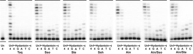
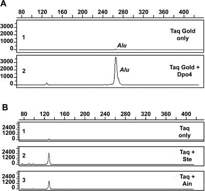
For many years, Taq polymerase has served as the stalwart enzyme in the PCR amplification of DNA. However, a major limitation of Taq is its inability to amplify damaged DNA, thereby restricting its usefulness in forensic applications. In contrast, Y-family DNA polymerases, such as Dpo4 from Sulfolobus solfataricus, can traverse a wide variety of DNA lesions. Here, we report the identification and characterization of five novel thermostable Dpo4-like enzymes from Acidianus infernus, Sulfolobus shibatae, Sulfolobus tengchongensis, Stygiolobus azoricus and Sulfurisphaera ohwakuensis, as well as two recombinant chimeras that have enhanced enzymatic properties compared with the naturally occurring polymerases. The Dpo4-like polymerases are moderately processive, can substitute for Taq in PCR and can bypass DNA lesions that normally block Taq. Such properties make the Dpo4-like enzymes ideally suited for the PCR amplification of damaged DNA samples. Indeed, by using a blend of Taq and Dpo4-like enzymes, we obtained a PCR amplicon from ultraviolet-irradiated DNA that was largely unamplifyable with Taq alone. The inclusion of thermostable Dpo4-like polymerases in PCRs, therefore, augments the recovery and analysis of lesion-containing DNA samples, such as those commonly found in forensic or ancient DNA molecular applications.


Figures

Figure 1
Figure 1
Identification, cloning, and phylogenetic analysis of novel Dpo4-like orthologs. (A) Illustration of the cloning strategy for identification and isolation of full-length dpo4-like orthologs. In some cases, degenerate PCR primers based on the Dpo4 and Dbh amino acid sequences were used to amplify a section a dpo4-like ortholog. Subsequently, degenerate PCR primers based on the amino acid sequences of the conserved upstream ribokinase (DTTGAGD) and downstream hypothetical (YEDVEGG) genes in combination with dpo4 ortholog gene-specific primers were used to amplify the beginning and the ending of each gene. In the case of the Ste dpo4-like gene, degenerate PCR primers based on the Dpo4 and Dbh amino acid sequences were used instead of gene-specific primers to isolate the ends of the gene. Once the sequence of the 5′ and 3′ of each gene was identified, primers were designed to amplify and clone the full-length genes. (B) Unrooted phylogenetic tree of Dpo4-like polymerases. A protein alignment was performed using the Clustal X program (version 1.62b) (27). This protein alignment was used to calculate the tree using the Draw N-J Tree algorithm. The unrooted tree was drawn from the calculated tree using the TreeView program (version 1.6) (28). The derivation of each protein is designated as follows: S.acidocaldarius (SacDbh), S.solfataricus Dpo4 (SsoDpo4), S.tokodaii (StoDpo4), S.shibatae (SshDpo4), S.tengchongensis (SteDpo4), A.infernus (AinDpo4), S.azoricus (SazDpo4) and S.ohwakuensis (SohDpo4).
Figure 2
Figure 2
Purification and processivity of Dpo4-like proteins. (A) Scheme for the purification of Dpo4-like proteins. Whole-cell extracts (WC) were clarified and then heat treated at 70°C for 10 min. Denatured E.coli proteins were removed from the extracts by centrifugation (70). Subsequently, the soluble extracts were passed over a Hydroxyapatite Bio-Gel HTP Gel column (HAP) and then a SP Sepharose HP column (Sph). The Saz and Soh Dpo4-like proteins were purified over one additional column, a HiLoad 26/60 Superdex 75 column (data not shown). M designates the Benchmark prestained protein size marker (Invitrogen). (B) M13mp18 primer extension reactions using the previously identified S.solfataricus Dpo4 (Sso) and the five newly identified polymerases; S.tengchongensis (Ste), S.shibatae (Ssh), A.infernus (Ain), S.azoricus (Saz) and S.ohwakuensis (Soh). Primer extension of primer M13HTP (5′-CCT TAG AAT CCT TGA AAA CAT AGC GA-3′) annealed to M13mp18 from base pairs 4101 to 4126 was performed at 60°C for 5 min utilizing 10 nM of the M13HTP/M13mp18 primer-template and increasing concentrations of the Dpo4-like enzymes; from left to right 0.2, 2.0 and 20 nM. Replication products were separated on 12%/8 M urea polyacrylamide gels and visualized by PhosphorImager analysis. Primer location and number of nucleotides added are indicated to the left of the gel. (C) M13mp18 primer extension reactions using the ‘little finger’ domain chimeric polymerases; A.infernus/S.solfataricus (Ain/Sso) and A.infernus/S.tengchongensis (Ain/Ste).
Figure 3
Figure 3
PCR amplification of the 1.1 kb S.tengchongensis Dpo4-like gene by Dpo4-like polymerases. A total of 2.5 U of Taq or 200 nM (400 nM of Ssh) of each Dpo4-like polymerase was added to the PCRs. Two microliters of the 50 µl PCRs were loaded onto a 0.9% agarose gel and the PCR products separated by electrophoresis. The enzymes employed in the PCRs are specified above each lane. The position of the 1.1 kb PCR product is indicated on the left. L and 100 designates the lambda BstE II and 100 bp markers, respectively.
Figure 4
Figure 4
Primer extension on CPD and abasic site-containing templates. Primer SSHTP2 (5′-GCG GTG TAG AGA CGA GTG CGG AG-3′) was annealed to the undamaged template, HTU50 (Un) (5′-CTC TCA CAA GCA GCC AGG CAT TCT CCG CAC TCG TCT CTA CAC CGC TCC GC-3′); the CPD-containing template, HMTT50 (CPD) (5′-CTC TCA CAA GCA GCC AGG CAT TCT CCG CAC TCG TCT CTA CAC CGC TCC GC-3′) (where the T-T CPD is underlined); or the abasic site-containing template, HTX50 (Abasic) (5′-CTC TCA CAA GCA GCC AGG CAT XCT CCG CAC TCG TCT CTA CAC CGC TCC GC-3′), (where X denotes the position of the abasic site). Primer extension assays utilized 10 nM of the three different primer/templates. The reactions containing the undamaged template (HTU50) was incubated at 60°C for 3 min, whereas the reactions containing the CPD (HMTT50) and abasic site (HTX50) templates were incubated at 60°C for 10 min. The concentrations of each enzyme utilized for the undamaged template (HTU50) reactions is as follows: Taq, 0.0025 U; Sso, 0.1 nM; Ste, 0.33 nM; Ssh, 0.4 nM; Ain, 0.2 nM; Ain/Sso, 0.5 nM; Ain/Ste, 0.5 nM. The concentrations of each enzyme utilized for the CPD-containing template (HMTT50) reactions was 50 times higher than that used for the undamaged template and is as follows: Taq, 0.125 U; Sso, 5 nM; Ste, 16.5 nM; Ssh, 20 nM; Ain, 10 nM; Ain/Sso, 25 nM; Ain/Ste, 25 nM. The concentrations of each enzyme utilized for the abasic site-containing template (HMTT50) reactions was five times higher than used for the undamaged template and is as follows: Taq, 0.0125 U; Sso, 0.5 nM; Ste, 1.65 nM; Ssh, 2 nM; Ain, 1 nM; Ain/Sso, 2.5 nM; Ain/Ste, 2.5 nM. Reactions were initiated by the addition of 100 µM of all four dNTPs (4) or 100 µM of individual dNTPs (G, A, T, C) (indicated below each lane). Replication products were separated on12%/8 M urea polyacrylamide gels and visualized by PhosphorImager analysis.
Figure 5
Figure 5
Primer extension on the hydantoin-containing template. Primer SSHydP (5′-AGA TCA GTC ACG-3′) was annealed to the undamaged template, HydU22 (Un) (5′-CAC TTC GGA TCG TGA CTG ATC T-3′), and the 5-hydroxy-5-methylhydantoin-containing template, (5′-CAC TTC GGA HCG TGA CTG ATC T-3′), (where H indicates the position of the Hydantoin). Primer extension assays utilized 10 nM of these primer/templates and the reactions were incubated at 37°C for 5 min. The concentrations of each enzyme utilized for the undamaged template (HTU50) and hydantoin-containing template reactions is as follows: Taq, 0.0083 U; Sso, 0.33 nM; Ste, 0.75 nM; Ssh, 1 nM; Ain, 0.5 nM; Ain/Sso, 1.25 nM; Ain/Ste, 1.25 nM. Reactions were initiated by the addition of 100 µM of all four dNTPs (4) or 100 µM of individual dNTPs (G, A, T, C) (indicated below each lane). Replication products were separated on 18%/8 M urea polyacrylamide gels and visualized by PhosphorImager analysis.
Figure 6
Figure 6
PCR amplification of UV-damaged DNA. (A) Human genomic DNA (K562) was damaged by exposure to UVC at a flux rate of 0.197 J/cm2/min. Two nanograms of damaged genomic DNA were amplified with primers specific for the Major and Precise Alu subfamilies (21). The PCR contained, inter alia, 2.5 U Taq Gold DNA Polymerase, 20 pmol each of the forward and reverse primers and, if applicable, 100 nM Dpo4. The forward primer was labeled at its 5′ end with the reactive fluorescent dye 6-FAM. Amplified fragments were separated by capillary electrophoresis and detected by laser-induced fluorescence of the incorporated dye-labeled primer. The electropherograms depict Alu element amplification products of the UVC damaged genomic substrate with Taq Gold DNA polymerase alone (1) or with a cocktail of Taq DNA polymerase and 100 nM Dpo4 (2). (B) Experimental details are the same as (A) except that the DNA was exposed to UVC at the same flux rate for 30 min and PCR was performed at 85°C, as noted in Materials and Methods. Results show the Alu element products obtained with AmpliTaq DNA polymerase alone (1) or with a cocktail of Taq DNA polymerase and 100 nM Ste (2) or Ain (3).

References

    1. Ohmori H., Friedberg E.C., Fuchs R.P.P., Goodman M.F., Hanaoka F., Hinkle D., Kunkel T.A., Lawrence C.W., Livneh Z., Nohmi T., et al. The Y-family of DNA polymerases. Mol. Cell. 2001;8:7–8. - PubMed
    1. Kulaeva O.I., Koonin E.V., McDonald J.P., Randall S.K., Rabinovich N., Connaughton J.F., Levine A.S., Woodgate R. Identification of a DinB/UmuC homolog in the archeon Sulfolobus solfataricus. Mutat. Res. 1996;357:245–253. - PubMed
    1. Boudsocq F., Iwai S., Hanaoka F., Woodgate R. Sulfolobus solfataricus P2 DNA polymerase IV (Dpo4): an archaeal DNA polymerase with lesion-bypass properties akin to eukaryotic polη. Nucleic Acids Res. 2001;29:4607–4616. - PMC - PubMed
    1. Gruz P., Pisani F.M., Shimizu M., Yamada M., Hayashi I., Morikawa K., Nohmi T. Synthetic activity of Sso DNA polymerase Y1, an archaeal DinB-like DNA polymerase, is stimulated by processivity factors proliferating cell nuclear antigen and replication factor C. J. Biol. Chem. 2001;276:47394–47401. - PubMed
    1. Potapova O., Grindley N.D., Joyce C.M. The mutational specificity of the dbh lesion bypass polymerase and its implications. J. Biol. Chem. 2002;277:28157–28166. - PubMed

Publication types

MeSH terms