Common dynamical signatures of familial amyotrophic lateral sclerosis-associated structurally diverse Cu, Zn superoxide dismutase mutants
- PMID: 16488975
- PMCID: PMC1413921
- DOI: 10.1073/pnas.0511266103
Common dynamical signatures of familial amyotrophic lateral sclerosis-associated structurally diverse Cu, Zn superoxide dismutase mutants
Abstract
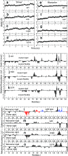
More than 100 structurally diverse point mutations leading to aggregation in the dimeric enzyme Cu, Zn superoxide dismutase (SOD1) are implicated in familial amyotrophic lateral sclerosis (FALS). Although SOD1 dimer dissociation is a known requirement for its aggregation, the common structural basis for diverse FALS mutations resulting in aggregation is not fully understood. In molecular dynamics simulations of wild-type SOD1 and three structurally diverse FALS mutants (A4V, G37R, and H46R), we find that a common effect of mutations on SOD1 dimer is the mutation-induced disruption of dynamic coupling between monomers. In the wild-type dimer, the principal coupled motion corresponds to a "breathing motion" of the monomers around an axis parallel to the dimer interface, and an opening-closing motion of the distal metal-binding loops. These coupled motions are disrupted in all three mutants independent of the mutation location. Loss of coupled motions in mutant dimers occurs with increased disruption of a key stabilizing structural element (the beta-plug) leading to the de-protection of edge strands. To rationalize disruption of coupling, which is independent of the effect of the mutation on global SOD1 stability, we analyze the residue-residue interaction network formed in SOD1. We find that the dimer interface and metal-binding loops, both involved in coupled motions, are regions of high connectivity in the network. Our results suggest that independent of the effect on protein stability, altered protein dynamics, due to long-range communication within its structure, may underlie the aggregation of mutant SOD1 in FALS.
Conflict of interest statement
Conflict of interest statement: No conflicts declared.
Figures


Similar articles
-
Unveiling the unfolding pathway of FALS associated G37R SOD1 mutant: a computational study.Mol Biosyst. 2010 Jun;6(6):1032-9. doi: 10.1039/b918662j. Epub 2010 Feb 23. Mol Biosyst. 2010. PMID: 20485746
-
FALS mutations in Cu, Zn superoxide dismutase destabilize the dimer and increase dimer dissociation propensity: a large-scale thermodynamic analysis.Amyloid. 2006 Dec;13(4):226-35. doi: 10.1080/13506120600960486. Amyloid. 2006. PMID: 17107883
-
An intersubunit disulfide bond prevents in vitro aggregation of a superoxide dismutase-1 mutant linked to familial amytrophic lateral sclerosis.Biochemistry. 2004 May 4;43(17):4899-905. doi: 10.1021/bi030246r. Biochemistry. 2004. PMID: 15109247
-
Structure, folding, and misfolding of Cu,Zn superoxide dismutase in amyotrophic lateral sclerosis.Biochim Biophys Acta. 2006 Nov-Dec;1762(11-12):1025-37. doi: 10.1016/j.bbadis.2006.05.004. Epub 2006 May 22. Biochim Biophys Acta. 2006. PMID: 16814528 Review.
-
[Familial amyotrophic lateral sclerosis and mutations in the Cu/Zn superoxide dismutase gene].Rinsho Shinkeigaku. 1995 Dec;35(12):1546-8. Rinsho Shinkeigaku. 1995. PMID: 8752459 Review. Japanese.
Cited by
-
Early steps in thermal unfolding of superoxide dismutase 1 are similar to the conformational changes associated with the ALS-associated A4V mutation.Protein Eng Des Sel. 2013 Aug;26(8):503-13. doi: 10.1093/protein/gzt030. Epub 2013 Jun 19. Protein Eng Des Sel. 2013. PMID: 23784844 Free PMC article.
-
Modifications of superoxide dismutase (SOD1) in human erythrocytes: a possible role in amyotrophic lateral sclerosis.J Biol Chem. 2009 May 15;284(20):13940-13947. doi: 10.1074/jbc.M809687200. Epub 2009 Mar 19. J Biol Chem. 2009. PMID: 19299510 Free PMC article.
-
Conformational specificity of the C4F6 SOD1 antibody; low frequency of reactivity in sporadic ALS cases.Acta Neuropathol Commun. 2014 May 14;2:55. doi: 10.1186/2051-5960-2-55. Acta Neuropathol Commun. 2014. PMID: 24887207 Free PMC article.
-
Molecular Mechanisms of Aggregation of Canine SOD1 E40K Amyloidogenic Mutant Protein.Molecules. 2022 Dec 24;28(1):156. doi: 10.3390/molecules28010156. Molecules. 2022. PMID: 36615350 Free PMC article.
-
Improving binding specificity of pharmacological chaperones that target mutant superoxide dismutase-1 linked to familial amyotrophic lateral sclerosis using computational methods.J Med Chem. 2010 Apr 8;53(7):2709-18. doi: 10.1021/jm901062p. J Med Chem. 2010. PMID: 20232802 Free PMC article.
References
-
- Valentine J. S., Doucette P. A., Potter S. Z. Annu. Rev. Biochem. 2005;74:563–593. - PubMed
-
- Rosen D. R., Siddique T., Patterson D., Figlewicz D. A., Sapp P., Hentati A., Donaldson D., Goto J., Oregan J. P., Deng H. X., et al. Nature. 1993;362:59–62. - PubMed
-
- Gurney M. E., Pu H. F., Chiu A. Y., Dalcanto M. C., Polchow C. Y., Alexander D. D., Caliendo J., Hentati A., Kwon Y. W., Deng H. X., et al. Science. 1994;264:1772–1775. - PubMed
-
- Gaudette M., Hirano M., Siddique T. Amyotrophic Lateral Scler. Other Motor Neuron Disorders. 2000;1:83–89. - PubMed
-
- Cleveland D. W., Rothstein J. D. Nat. Rev. Neurosci. 2001;2:806–819. - PubMed
Publication types
MeSH terms
Substances
LinkOut - more resources
Full Text Sources
Medical
Miscellaneous

