

Self-assembly of synthetic collagen triple helices
- PMID: 16488977
- PMCID: PMC1413887
- DOI: 10.1073/pnas.0508783103
Self-assembly of synthetic collagen triple helices
Abstract
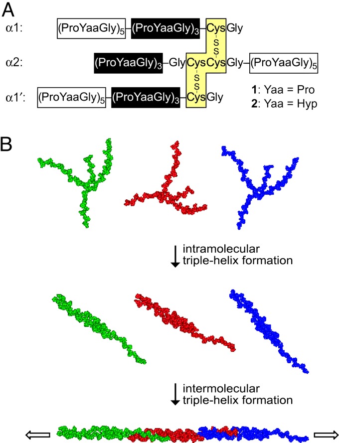
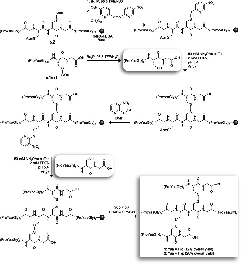
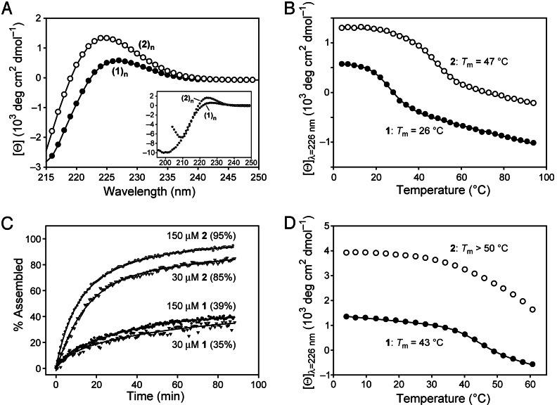
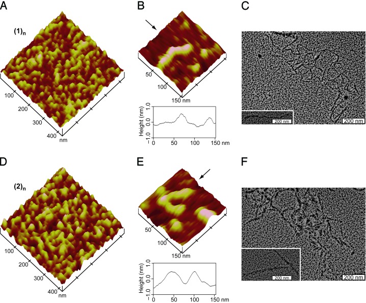
Collagen is the most abundant protein in animals and the major component of connective tissues. Although collagen isolated from natural sources has long served as the basis for some biomaterials, natural collagen is difficult to modify and can engender pathogenic and immunological side effects. Collagen comprises a helix of three strands. Triple helices derived from synthetic peptides are much shorter (<10 nm) than natural collagen (approximately 300 nm), limiting their utility. Here, we describe the synthesis of short collagen fragments in which the three strands are held in a staggered array by disulfide bonds. Data from CD spectroscopy, dynamic light scattering, analytical ultracentrifugation, atomic force microscopy, and transmission electron microscopy indicate that these "sticky-ended" fragments self-assemble via intermolecular triple-helix formation. The resulting fibrils resemble natural collagen, and some are longer (>400 nm) than any known collagen. We anticipate that our self-assembly strategy can provide synthetic collagen-mimetic materials for a variety of applications.
Conflict of interest statement
Conflict of interest statement: No conflicts declared.
Figures
References
-
- Ramshaw J. A. M., Werkmeister J. A., Glattauer V. Biotechnol. Genet. Eng. Rev. 1996;13:335–382. - PubMed
-
- Lee C. H., Singla A., Lee Y. Int. J. Pharm. 2001;221:1–22. - PubMed
-
- Cooperman L., Michaeli D. J. Am. Acad. Dermatol. 1984;10:638–646. - PubMed
-
- Sakaguchi M., Hori H., Hattori S., Irie S., Imai A., Yanagida M., Miyazawa H., Toda M., Inouye S. J. Allergy Clin. Immunol. 1999;104:695–699. - PubMed
-
- Lynn A. K., Yannas I. V., Bonfield W. J. Biomed. Mater. Res. B. 2004;71:343–354. - PubMed
Publication types
MeSH terms
Substances
Grants and funding
LinkOut - more resources
Full Text Sources
Other Literature Sources
Molecular Biology Databases
