. 2006 Feb 28;103(9):3375-80.
doi: 10.1073/pnas.0507749103. Epub 2006 Feb 17.

Identification of a domain in Yersinia virulence factor YadA that is crucial for extracellular matrix-specific cell adhesion and uptake

Affiliations

Identification of a domain in Yersinia virulence factor YadA that is crucial for extracellular matrix-specific cell adhesion and uptake

Tanja Heise et al. Proc Natl Acad Sci U S A. .

Abstract

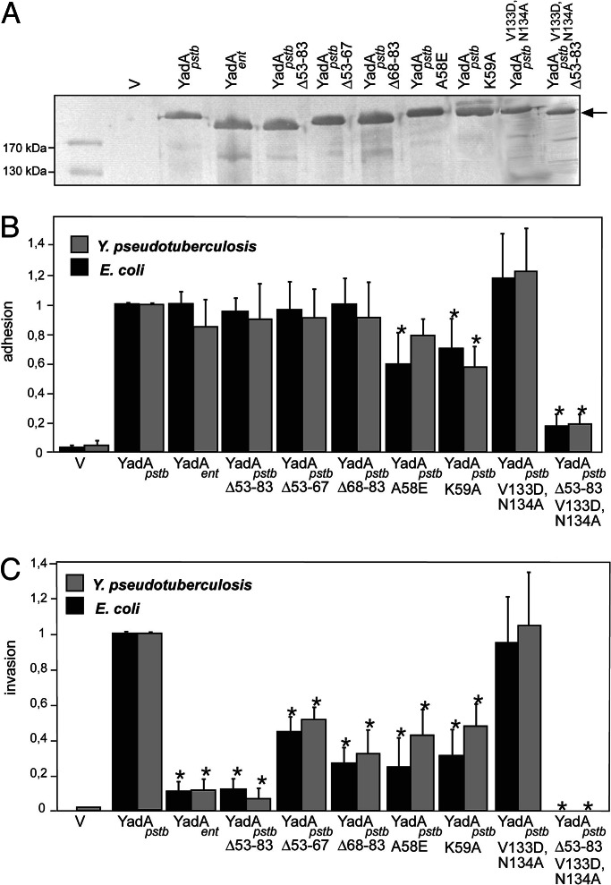
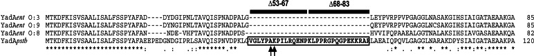
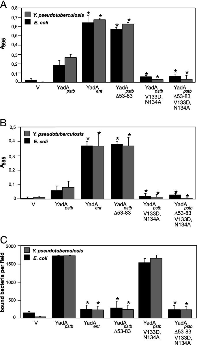
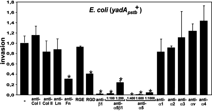
For many pathogens, cell adhesion factors are critical virulence determinants. Enteropathogenic Yersinia species express the afimbrial adhesin YadA, the prototype of a class of homotrimeric outer membrane adhesins, which mediates adherence to host cells by binding to extracellular matrix components. In this study, we demonstrate that different pathogenic functions are attributable to highly homologous YadA proteins. YadA of Yersinia pseudotuberculosis (YadA(pstb)) and Yersinia enterocolitica (YadA(ent)) exhibit fundamental differences in their specificity of extracellular matrix substrate binding, they cause dissimilar bacterial aggregation behaviors, and YadA(pstb), but not YadA(ent), promotes efficient uptake into human cells. Evidence is presented here that a unique N-terminal amino acid sequence of YadA(pstb), which is absent in YadA(ent), acts as an "uptake domain" by mediating tight binding to fibronectin bound on alpha(5)beta(1) integrin receptors, which are crucial for initiating the entry process. Deleting this motif in YadA(pstb) generated all features of the YadA(ent) protein, i.e., the molecule lost its adhesiveness to fibronectin and its invasiveness, but gained adhesion potential to collagen and laminin. Loss of the "uptake region" also attenuated host tissue colonization by Y. pseudotuberculosis during oral infections of mice, demonstrating that this motif plays a crucial role in defining pathogen-host cell interaction and pathogenesis. We conclude that even small variations in adhesion factors can provoke major differences in the virulence properties of related pathogens.


Conflict of interest statement

Conflict of interest statement: No conflicts declared.

Figures

Fig. 1.
Fig. 1.
Outer membrane incorporation, adhesion, and invasion properties of the YadA proteins. (A) Immunoblot analysis demonstrates that comparable amounts of multimeric YadA derivatives (180–200 kDa) are expressed in the outer membrane of Yersinia strain YP31. V, empty vector. Cell adhesion (B) and cell invasion (C) of YadA-expressing E. coli and Y. pseudotuberculosis YP31 recombinant strains are documented. Approximately 106 bacteria were used to challenge 5 × 10⁴ HEp-2 cells. Total numbers of adherent and intracellular bacteria are expressed relative to the adhesion and invasion rate of E. coli and Y. pseudotuberculosis YP31 harboring yadApstb, defined as 1.0. Each value represents the mean of at least three independent assays done in triplicate. Data were analyzed by the Student t test. ∗, significantly different from YadApstb-expressing bacteria (P < 0.05).
Fig. 2.
Fig. 2.
clustalw alignment of the head domain of YadA of Y. enterocolitica Serotype O:3 (YadAent O:3), O:9 (YadAent O:9) and O:8 (YadAent O:8), and Y. pseudotuberculosis Type III (YadApstb). Asterisks indicate amino acid identity, and dots indicate amino acid similarity. The additional sequence in YadA of Y. pseudotuberculosis, deleted in the YadApstbΔ53–83 protein, is indicated with two adjacent black boxes, which also illustrate the portions of the smaller deletions in YadApstbΔ53–67 and YadApstbΔ68–83. Exchanged amino acids A58E, K59A in the head domain of the YadApstb mutant derivatives are indicated by arrows.
Fig. 3.
Fig. 3.
Different binding properties of YadA-expressing E. coli and Y. pseudotuberculosis YP31 strains to ECM molecules. Plastic wells were coated with collagen type I (A) or laminin (B). Bound bacteria were stained, and absorbance was measured at 595 nm. Expression of YadApstb results in a moderate interaction, whereas bacteria expressing YadAent and YadApstbΔ53–83 exhibit a strong binding capacity to collagen and laminin. Data represent the mean ± SD of three independent experiments done in duplicate. For quantitative analysis of adherence to fibronectin (C), glass coverslips were coated with cellular fibronectin and bound bacteria were counted in a defined area of a microscopic field of ≈4 × 103 μm2. The means and SD of 30 calculated fields are shown. Data were analyzed by the Student t test. ∗, significantly different from YadApstb-expressing bacteria (P < 0.05).
Fig. 4.
Fig. 4.
Inhibition of YadA-promoted invasion by blockage of fibronectin and the fibronectin-binding class of β1 integrins. HEp-2 cells were blocked with different antibodies directed against ECM molecules (Col, collagen; Lm, Laminin; Fn, fibronectin), GRGDSP/GRGESP peptides, and with antibodies to different α-subunits of β1 integrins before infection with YadA-expressing bacteria. Approximately 106 bacteria were used to challenge 5 × 10⁴ cells, which were incubated for 1 h at 37°C to determine cell uptake efficiency. Only blockage of fibronectin and α5β1 integrins reduced YadA uptake. The proportionate reduction in invasion is expressed relative to the invasion rate promoted by the YadApstb WT protein, defined as 1.0. Each value represents the mean of three independent assays done in triplicate. Data were analyzed by the Student t test. ∗, P < 0.05 relative to YadApstb without antibodies/peptides (−).

Similar articles

Cited by

References

    1. Bottone E. J. Clin. Microbiol. Rev. 1997;10:257–276. - PMC - PubMed
    1. Pepe J. C., Wachtel M. R., Wagar E., Miller V. L. Infect. Immun. 1995;63:4837–4848. - PMC - PubMed
    1. Marra A., Isberg R. R. Infect. Immun. 1997;65:3412–3421. - PMC - PubMed
    1. Leong J. M., Fournier R. S., Isberg R. R. EMBO J. 1990;9:1979–1989. - PMC - PubMed
    1. Clark M. A., Hirst B. H., Jepson M. A. Infect. Immun. 1998;66:1237–1243. - PMC - PubMed

Publication types

MeSH terms