. 2006 Feb 28;45(8):2524-36.
doi: 10.1021/bi052475e.

Mitochondrial matrix phosphoproteome: effect of extra mitochondrial calcium

Affiliations

Mitochondrial matrix phosphoproteome: effect of extra mitochondrial calcium

Rachel K Hopper et al. Biochemistry. .

Abstract

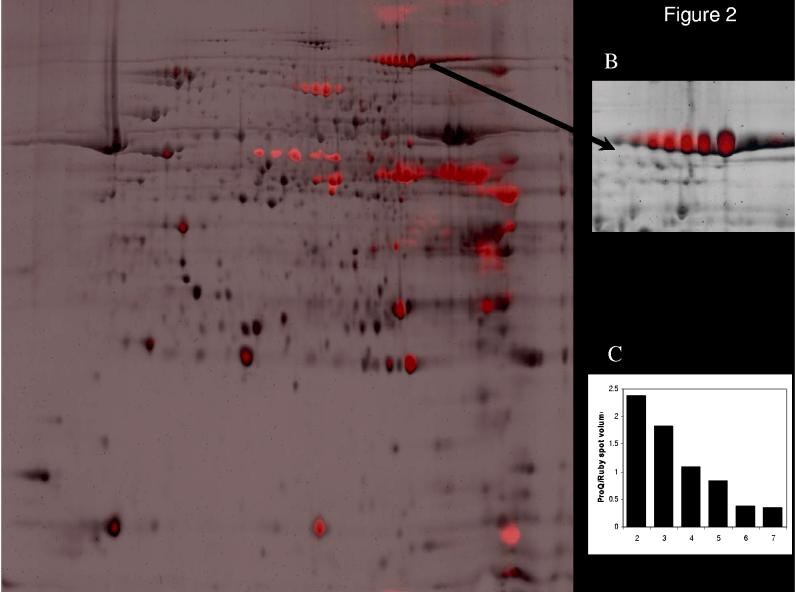
Post-translational modification of mitochondrial proteins by phosphorylation or dephosphorylation plays an essential role in numerous cell signaling pathways involved in regulating energy metabolism and in mitochondrion-induced apoptosis. Here we present a phosphoproteomic screen of the mitochondrial matrix proteins and begin to establish the protein phosphorylations acutely associated with calcium ions (Ca(2+)) signaling in porcine heart mitochondria. Forty-five phosphorylated proteins were detected by gel electrophoresis-mass spectrometry of Pro-Q Diamond staining, while many more Pro-Q Diamond-stained proteins evaded mass spectrometry detection. Time-dependent (32)P incorporation in intact mitochondria confirmed the extensive matrix protein phosphoryation and revealed the dynamic nature of this process. Classes of proteins that were detected included all of the mitochondrial respiratory chain complexes, as well as enzymes involved in intermediary metabolism, such as pyruvate dehydrogenase (PDH), citrate synthase, and acyl-CoA dehydrogenases. These data demonstrate that the phosphoproteome of the mitochondrial matrix is extensive and dynamic. Ca(2+) has previously been shown to activate various dehydrogenases, promote the generation of reactive oxygen species (ROS), and initiate apoptosis via cytochrome c release. To evaluate the Ca(2+) signaling network, the effects of a Ca(2+) challenge sufficient to release cytochrome c were evaluated on the mitochondrial phosphoproteome. Novel Ca(2+)-induced dephosphorylation was observed in manganese superoxide dismutase (MnSOD) as well as the previously characterized PDH. A Ca(2+) dose-dependent dephosphorylation of MnSOD was associated with an approximately 2-fold maximum increase in activity; neither the dephosphorylation nor activity changes were induced by ROS production in the absence of Ca(2+). These data demonstrate the use of a phosphoproteome screen in determining mitochondrial signaling pathways and reveal new pathways for Ca(2+) modification of mitochondrial function at the level of MnSOD.


Figures

Fig. 1
Fig. 1
Two-dimensional gel electrophoresis and staining of the phosphoproteome of porcine heart mitochondria with Pro-Q Diamond phosphoprotein gel stain. Proteins are separated by isoelectric point (pI), from pH ∼4-9 along the horizontal axis, and by molecular weight, from ∼100 to 10 kD, vertically. Numbers refer to the protein identifications presented in Table 1. Not all Pro-Q Diamond-stained proteins were identified due to not reaching statistical significance in the mass spectrometry analysis.
Fig. 2
Fig. 2
Overlay of Sypro Ruby total protein (black) and Pro-Q Diamond phosphoprotein (red) staining of mitochondrial proteome in the absence of Ca2+ (A). The relative amplitude of the two channels was arbitrarily set. The majority of proteins detected by Sypro Ruby were not detected with Pro-Q Diamond resulting in a predominance of pure black spots. Proteins heavily labeled with Pro-Q Diamond appear red with essentially no Sypro Ruby signal (for example PDH, spots 27-33). We used the ratio of the Sypro Ruby and Pro-Q Diamond signals as a quantitative method for determining the degree of protein phosphorylation. As a control for this approach, the multiple phosphorylation states of aconitase (spots 2-7) were evaluated in panels B and C. The enhanced phosphorylation of aconitase is associated with an acid shift in its isoelectric focusing pH, taking the ratio of the Sypro Ruby stain and Pro-Q Diamond revealed a quantitative relationship between this ratio and isoelectic focus for this single protein.
Fig. 3
Fig. 3
Two-dimensional gel electrophoresis and staining of the phosphoproteome of porcine heart mitochondria with P32-PO4. Proteins are separated by isoelectric point (pI), from pH ∼4-9 along the horizontal axis, and by molecular weight, from ∼100 to 10 kD, vertically. A) Autoradiogram of gel. The MnSOD-Rieske iron sulfur region is expanded in this and all other panels at optimal contrast/brightness. B) Coomassie stain of same gel. C) Color overlay of autoradiogram (red) and Coomassie stain (green). Amplitude of both gels was arbitrarily set.
Fig. 4
Fig. 4
Two-dimensional gel electrophoresis and staining of the phosphoproteome of porcine heart mitochondria with 32P PO4. Proteins are separated by isoelectric point (pI), from pH ∼4-9 along the horizontal axis, and by molecular weight, from ∼100 to 10 kD, vertically. The mitochondria were harvested after incubation with 32P-PO4 for either 5 minutes (A), 20 minutes (B) or 20 minutes then 5 minutes with dinitrophenol, a mitochondrial uncoupler (C). The incubation conditions are outlined in the Methods section.
Fig. 5
Fig. 5
Effect of Ca2+ on PDH phosphorylation and activity. Representative images of gels stained with Pro-Q Diamond indicate the degree of PDH E1α phosphorylation of individual proteins under conditions of zero (top panel) and high free Ca2+ (bottom panel) (A). Multiple protein spots of pyruvate dehydrogenase E1 alpha subunit stain with Pro-Q Diamond more intensely under control conditions than under high Ca2+ conditions. The degree of phosphorylation under each condition was calculated as the ratio of intensity of Pro-Q staining for each spot to the total Sypro Ruby spot volume for that gel to normalize for any difference in total protein loaded in the gel and is given as the mean ± S.E.M. (B). Because these proteins are highly phosphorylated but not abundant, matching spots from Pro-Q Diamond to Sypro Ruby images was difficult and therefore total spot volume of the gel was used to normalize to amount of protein. PDH enzyme activity increased in the presence of high Ca2+ relative to control conditions (C).
Fig. 6
Fig. 6
The effect of Ca2+ on MnSOD phosphorylation and activity. A) MnSOD also showed less intense staining with Pro-Q Diamond under high Ca2+ conditions (bottom panel) compared to control (top panel). Quantification of the degree of phosphorylation under each condition was determined by the intensity of Pro-Q Diamond staining normalized to the corresponding Sypro Ruby intensity for that MnSOD spot (B). The activity of MnSOD normalized to cyt a content under control and high Ca2+ conditions shows increased enzyme activity with the addition of Ca2+ (C). This increase is dependent on Ca2+ concentration. The dose-response curve of MnSOD activity over Ca2+ conditions ranging from 0 to 100 μM free Ca2+, expressed as the percent activity under control conditions, show that the K50 is ∼10μM (D).
Fig. 7
Fig. 7
The effects of matrix ROS production on MnSOD phosphorylation and activity. Rate of H2O2 production per minute, normalized to cyt a content, shows that treatment of mitochondria with rotenone and succinate (R/S) increased rate of H2O2 production significantly over control levels, similar to the increase induced by the high Ca2+ conditions (A). Cyt c is released from mitochondria in the presence of high Ca2+, but not with R/S, indicating that the increased H2O2 production does not induce apoptosis (B). MnSOD spots in gels of mitochondria exposed to R/S show no change in Pro-Q Diamond staining intensity relative to control (C). MnSOD activity shows no significant difference under control and R/S conditions (D).
Fig. 8.
Fig. 8.
The [Ca2+] dose dependence of PDH and MnSOD phosphorylation Experiments were conducted under identical conditions as in Figures 5 and 6 with the free [Ca2+] of 0, 0.6, 40 and 100 μM.

References

    1. Gray MW. The endosymbiont hypothesis revisited. Int. Rev. Cytol. 1992;141:233–357. - PubMed
    1. Cavalier-Smith T. The simultaneous symbiotic origin of mitochondria, chloroplasts, and microbodies. Ann. N. Y. Acad. Sci. 1987;503:55–71. - PubMed
    1. Westermann B, Neupert W. ‘Omics’ of the mitochondrion. Nat. Biotechnol. 2003;21:239–240. - PubMed
    1. Richly E, Chinnery PF, Leister D. Evolutionary diversification of mitochondrial proteomes: implications for human disease. Trends Genet. 2003;19:356–362. - PubMed
    1. Taylor SW, Fahy E, Zhang B, Glenn GM, Warnock DE, Wiley S, Murphy AN, Gaucher SP, Capaldi RA, Gibson BW, Ghosh SS. Characterization of the human heart mitochondrial proteome. Nat. Biotechnol. 2003;21:281–286. - PubMed

Publication types

MeSH terms