. 1991 Jul 12;66(1):173-83.
doi: 10.1016/0092-8674(91)90149-s.

The trk tyrosine protein kinase mediates the mitogenic properties of nerve growth factor and neurotrophin-3

Affiliations

The trk tyrosine protein kinase mediates the mitogenic properties of nerve growth factor and neurotrophin-3

C Cordon-Cardo et al. Cell. .

Abstract

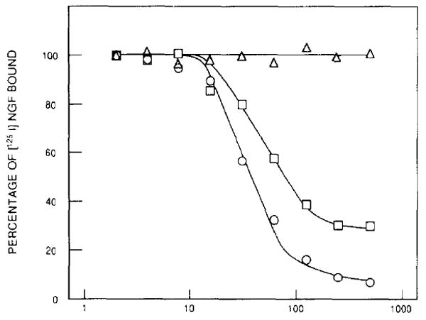
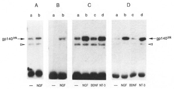
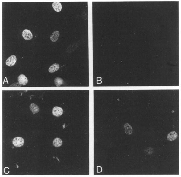
The product of the trk proto-oncogene encodes a receptor for nerve growth factor (NGF). Here we show that NGF is a powerful mitogen that can induce resting NIH 3T3 cells to enter S phase, grow in semisolid medium, and become morphologically transformed. These mitogenic effects are absolutely dependent on expression of gp140trk receptors, but do not require the presence of the previously described low affinity NGF receptor. gp140trk also serves as a receptor for the related factor neurotrophin-3 (NT-3), but not for brain-derived neurotrophic factor. Both NGF and NT-3 induce the rapid phosphorylation of gp140trk receptors and the transient expression of c-Fos proteins. However, NT-3 appears to elicit more limited mitogenic responses than NGF. These results indicate that the product of the trk proto-oncogene is sufficient to mediate signal transduction processes induced by NGF and NT-3, at least in proliferating cells.


Figures

Figure 1
Figure 1. Stimulation of [3H]Thymidine Incorporation by NGF
(A) Quiescent NIH 3T3, E25-48, and B35-41 cells were incubated with DMEM containing 5 μg/ml insulin (open bars), 20% calf serum (hatched bars), or 30 ng/ml NGF plus 5 μg/ml insulin (filled bars). Results are expressed as the mean of three independent experiments assayed in duplicate (+SEM). (B) E25-43 cells were stimulated by the indicated amounts of NGF either in the absence (open circles) or presence (filled circles) of a 10-fold molar excess of anti-NGF monoclonal antibody clone 27/21 (Boehringer Mannheim).
Figure 2
Figure 2. Induction of DNA Synthesis by NGF
Quiescent NIH 3T3 (A, C, E) and E2546 (B, D, F) cells were incubated in the presence of 100 μM Brdlhd plus either 20% calf serum (A, B), 5 μg/ml insulin (C, D), or 10 ng/ml NGF plus 5 μg/ml insulin (E, F). Cells were fixed after 24 hr and analyzed for immunofluorescence using a mouse anti-BrdUrd monoclonal antibody and donkey anti-mouse IgG conjugated with Texas red. Final magnification 250 x
Figure 3
Figure 3. Responsiveness to NGF Correlates with Expression of the trk Proto-Oncogene Product
E25-48 cells were assayed for BrdUrd incorporation as described in the legend to Figure 2 and the results photographically recorded. Microslides containing BrdUrd-stained E25-48 cells were then used to immunocytochemically visualize gp140trk expression using the avidin–biotin immunoperoxidase technique. (A) Preimmune rabbit serum. (B, C) Rabbit polyclonal anti-trk antiserum. (D) BrdUrd incorporation in the same field as shown in (C). Final magnifications: 270x (A, B) and 400x (C, D). Asterisks indicate cells without detectable gp140trk expression (C) and without BrdUrd incorporation (D).
Figure 4
Figure 4. NT-3 Competes with 125I-Labeled NGF for Binding to the trk Receptors Expressed in E25-48 Cells
E25-48 cells were preincubated with unlabeled NGF (circles), NT-3 (squares), or BDNF (triangles) at 4°C for 1 hr. 125I-labeled NGF was added to the cultures at a final concentration of 5 ng/ml, and the cells were incubated for an additional 2 hr. The percentage of 125I-labeled NGF bound to the cells was determined as described in Experimental Procedures. Results are expressed as the mean of duplicate samples.
Figure 5
Figure 5. NT-3 Induces DNA Synthesis in E25-48 Cells
The mitogenic activity of NT-3 was measured by immunofluorescence after BrdUrd incorporation. Cells were stimulated with DMEM containing 20% calf serum (A), 5 μg/ml insulin (B), or 50 ng/ml NT-3 plus 5 μg/ml insulin (C). Final magnification 250x.
Figure 6
Figure 6. NGF and NT-3 Stimulate Tyrosine Phosphorylation of the trk Proto-Oncogene Product
Quiescent E25-48 cells were incubated for 10 min at 37°C with DMEM alone (lanes a) or with DMEM containing 50 ng/ml NGF (lanes b), BDNF (lanes c), or NT-3 (lanes d). Cells were lysed in RIPAE buffer containing 0.5 mM sodium orthovanadate, immunoprecipitated with a rabbit polyclonal antibody elicited against a peptide corresponding to the carboxy-terminal domain of gp140trk (Martin-Zanca et al., 1989) and analyzed by 8% SDS–polyacrylamide gel electrophoresis. The electrophoresed samples were transferred to nitrocellulose filters and blotted with anti-trk antiserum (A) or anti-phosphotyrosine monoclonal antibody 4G10 (UBI) (B–D) as described in Experimental Procedures. (C) depicts tyrosine phosphorylation of gp140trk after stimulation with baculovirus-expressed and BDNF and NT-3, (D) after stimulation with COS cell–expressed BNDF and NT-3. The resulting nitrocellulose filters were exposed to Kodak X-Omat film for 8 hr at −70°C with the help of an intensifying screen.
Figure 7
Figure 7. NGF and NT-3 Induce Expression of c-Fos Protein in E25-48 Cells
Quiescent E25-48 cells were incubated at 37°C for 90 min in DMEM containing either 20% calf serum (A), 5 μg/ml insulin (B), 50 ng/ml NGF (C), or 50 ng/ml NT-3 (D). After fixation in methanol, immunofluorescence was performed using anti-c-Fos antibodies and anti-rabbit IgG conjugated with rhodamine. Final magnification 390x.
Figure 8
Figure 8. Kinetics of c-Fos Induction by NGF
Quiescent PC12 (A) and E25-48 (B) cells were stimulated with 100 ng/ml NGF for the indicated times in minutes, pulse labeled for 30 min with 400 μCi/ml [35S]methionine, and the cell lysates immunoprecipitated with rabbit polyclonal antic-Fos antibodies. lmmunoprecipitates were analyzed by 12% SDS–polyacrylamide gel electrophoresis. Gels were subjected to fluorography, dried, and exposed to Kodak X-Omat film for 3 days at −70°C with the help of an intensifying screen. Coelectrophoresed molecular weight markers include phosphorylase B (92,000) albumin (69,000), ovalbumin (46,000) and carbonic anhydrase (30,000).

References

    1. Alema S, Casalbore P, Agostini E, Tato F. Differentiation of PC12 phaeochromocytoma cells induced by v-src oncogene. Nature. 1985;316:557–559. - PubMed
    1. Aloe L, Levi-Montalcini R. Mast cells increase in tissues of neonatal rats injected with the nerve growth factor. Brain Res. 1977;133:358–366. - PubMed
    1. Barbacid M, Lamballe F, Pulido D, Klein R. The trk family of tyrosine protein kinase receptors. Biochim Biophys Acta Rev Cancer. 1991 in press. - PubMed
    1. Barde Y-A, Edgar D, Thoenen H. Purification of a new neurotrophic factor from mammalian brain. EMBO J. 1982;1:549–553. - PMC - PubMed
    1. Bar-Sagi D, Feramisco JR. Microinjection of the ras oncogene protein into PC12 cells induces morphological differentiation. Cell. 1985;42:841–848. - PubMed

Publication types

MeSH terms