Review
. 2006 Feb;6(2):136-47.
doi: 10.1038/nri1775.

Pathogen-endoplasmic-reticulum interactions: in through the out door

Affiliations
Review

Pathogen-endoplasmic-reticulum interactions: in through the out door

Craig R Roy et al. Nat Rev Immunol. 2006 Feb.

Abstract

A key determinant for the survival of intracellular pathogens is their ability to subvert the cellular processes of the host to establish a compartment that allows replication. Although most microorganisms internalized by host cells are efficiently cleared following fusion with lysosomes, many pathogens have evolved mechanisms to escape this degradation. In this Review, we provide insight into the molecular processes that are targeted by pathogens that interact with the endoplasmic reticulum and thereby subvert the immune response, ensure their survival intracellularly and cause disease. We also discuss how the endoplasmic reticulum 'strikes back' and controls microbial growth.


Conflict of interest statement

The authors declare no competing financial interests.

Figures
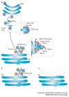
Figure 1
Figure 1. Biogenesis of the endoplasmic-reticulum–Golgi intermediate compartment and the Golgi from the endoplasmic reticulum.
a | Activation of SAR1 leads to the recruitment of the components of the coat complex COPII to endoplasmic reticulum (ER) membranes. COPII recruits cargo proteins that are to be delivered to other organelles and packages these into membrane tubules and vesicles. Activation of ADP-ribosylation factor 1 (ARF1) leads to recruitment of the components of the coat complex COPI to early secretory vesicles. On these membranes, exchange of the COPII coat for a COPI coat allows further protein sorting to occur and allows transport of cargo proteins away from the ER. Cells can then assemble a functional Golgi apparatus, and the secretory pathway is fully operational. b | In cells that express a particular mutant SAR1 protein (in which a histidine residue is replaced by a glycine residue at position 79) or a particular mutant ARF1 protein (in which a threonine residue is replaced by an asparagine residue at position 31), ER-exit sites are formed, but cargo is not transported away from the ER. c | In cells that express a different mutant SAR1 protein (in which a threonine residue is replaced by an asparagine residue at position 39), recruitment of the components of COPII to membranes is inhibited, and ER-exit sites do not form. ERGIC, endoplasmic-reticulum–Golgi intermediate compartment.
Figure 2
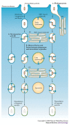
Figure 2. Models of phagosome maturation and cross-presentation.
a | In the first model, which is the classical model of phagocytosis and phagosome maturation, the plasma membrane invaginates and fuses, engulfing the extracellular particles and generating a phagosome. The phagosome then undergoes sequential interactions with both early and late endosomes, during which it acquires MHC class II molecules that are ready for peptide loading. b | In a second model of phagocytosis and phagosome maturation, in addition to interacting with the early and late endosomes, the plasma-membrane-derived phagosome undergoes several waves of fusion and fission with endoplasmic reticulum (ER)-derived vesicles. This could account for the delivery of MHC class I molecules and associated molecules to the phagosome and would facilitate cross-presentation of exogenously derived antigens. c | In a third model, recruitment of the ER to the site of phagocytosis provides MHC class I molecules and associated molecules at the beginning of the phagocytic process. The MHC class I molecules are retained in the phagosome until they are loaded with specific antigens that have been processed in the phagosome.
Figure 3
Figure 3. Pathogen–endoplasmic-reticulum interactions.
a | Viruses have evolved proteins that interfere with TAP (transporter associated with antigen processing) heterodimers in the lumen of the endoplasmic reticulum (ER): for example, the adenovirus protein E19, the human cytomegalovirus (CMV) proteins US3 and US6, and the herpes simplex virus (HSV) protein ICP47. The adenovirus protein E19 also interferes with retrograde transport, from the Golgi to the ER, and causes peptide-loaded MHC class I molecules to be returned to the ER. By contrast, several other proteins — such as the human CMV proteins US3 and US10, the hepatitis C virus (HCV) precursor protein non-structural protein 4A (NS4A)–NS4B, poliovirus protein 3A and coxsackievirus protein 3A — interfere with transport from the ER to the Golgi. b | Brucella-containing vacuoles require the activity of SAR1 for productive interactions with the ER. Cells that express a mutant SAR1 protein in which a threonine residue is replaced by an asparagine residue at position 39 (SAR1T39N) do not allow the replication of Brucella abortus. By contrast, Legionella pneumophila-containing vacuoles require the sequential activity of SAR1 and ADP-ribosylation factor 1 (ARF1) for productive interactions with the ER. Cells that express either a mutant SAR1 protein in which a histidine residue is replaced by a glycine residue at position 79 (SAR1H79G) or a mutant ARF1 protein in which a threonine residue is replaced by an asparagine residue at position 31 (ARF1T31N) do not allow replication of L. pneumophila. In the case of Toxoplasma gondii, the T. gondii proteins dense-granule protein 3 (GRA3) and rhoptry protein 2 (ROP2) might be involved in mediating interactions between the parasitophorous vacuole of T. gondii and the ER of the host. ERGIC, ER–Golgi intermediate compartment; β2m, β2-microglobulin.
Figure 4
Figure 4. The endoplasmic reticulum in host defence.
Although microbial pathogens have evolved mechanisms to subvert endoplasmic reticulum (ER) functions, the ER also has an important role in combating microorganisms. For example, under starvation conditions or following treatment with interferon-γ (IFNγ), host cells that are infected with Mycobacterium tuberculosis trigger autophagy, and the ER contributes to the formation of the autophagosome. Autophagosomes can then fuse with lysosomes, resulting in the destruction of M. tuberculosis trapped inside autophagosomes. In addition, infection with a virus, such as herpes simplex virus, induces the ER stress response, and exposure to IFNs induces the upregulation of expression of antimicrobial factors in the ER, including viperin and the p47 GTPases IGTP (IFNγ-induced GTPase) and IIGP1 (IFN-inducible GTPase 1).

Similar articles

Cited by

References

    1. Bonifacino JS, Glick BS. The mechanisms of vesicle budding and fusion. Cell. 2004;116:153–166. - PubMed
    1. Altan-Bonnet N, Sougrat R, Lippincott-Schwartz J. Molecular basis for Golgi maintenance and biogenesis. Curr. Opin. Cell Biol. 2004;16:364–372. - PubMed
    1. Barlowe C, et al. COPII: a membrane coat formed by Sec proteins that drive vesicle budding from the endoplasmic reticulum. Cell. 1994;77:895–907. - PubMed
    1. Roberg KJ, Crotwell M, Espenshade P, Gimeno R, Kaiser CA. LST1 is a SEC24 homologue used for selective export of the plasma membrane ATPase from the endoplasmic reticulum. J. Cell Biol. 1999;145:659–672. - PMC - PubMed
    1. Shimoni Y, et al. Lst1p and Sec24p cooperate in sorting of the plasma membrane ATPase into COPII vesicles in Saccharomyces cerevisiae. J. Cell Biol. 2000;151:973–984. - PMC - PubMed

Publication types