

Glucocorticoid-treated mice have localized changes in trabecular bone material properties and osteocyte lacunar size that are not observed in placebo-treated or estrogen-deficient mice
- PMID: 16491295
- PMCID: PMC1797152
- DOI: 10.1359/JBMR.051103
Glucocorticoid-treated mice have localized changes in trabecular bone material properties and osteocyte lacunar size that are not observed in placebo-treated or estrogen-deficient mice
Abstract
This study compares changes in bone microstructure in 6-month-old male GC-treated and female ovariectomized mice to their respective controls. In addition to a reduction in trabecular bone volume, GC treatment reduced bone mineral and elastic modulus of bone adjacent to osteocytes that was not observed in control mice nor estrogen-deficient mice. These microstructural changes in combination with the macrostructural changes could amplify the bone fragility in this metabolic bone disease.
Introduction: Patients with glucocorticoid (GC)-induced secondary osteoporosis tend to fracture at higher bone mineral densities than patients with postmenopausal osteoporosis. This suggests that GCs may alter bone material properties in addition to BMD and bone macrostructure.
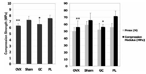
Materials and methods: Changes in trabecular bone structure, elastic modulus, and mineral to matrix ratio of the fifth lumbar vertebrae was assessed in prednisolone-treated mice and placebo-treated controls for comparison with estrogen-deficient mice and sham-operated controls. Compression testing of the third lumbar vertebrae was performed to assess whole bone strength.
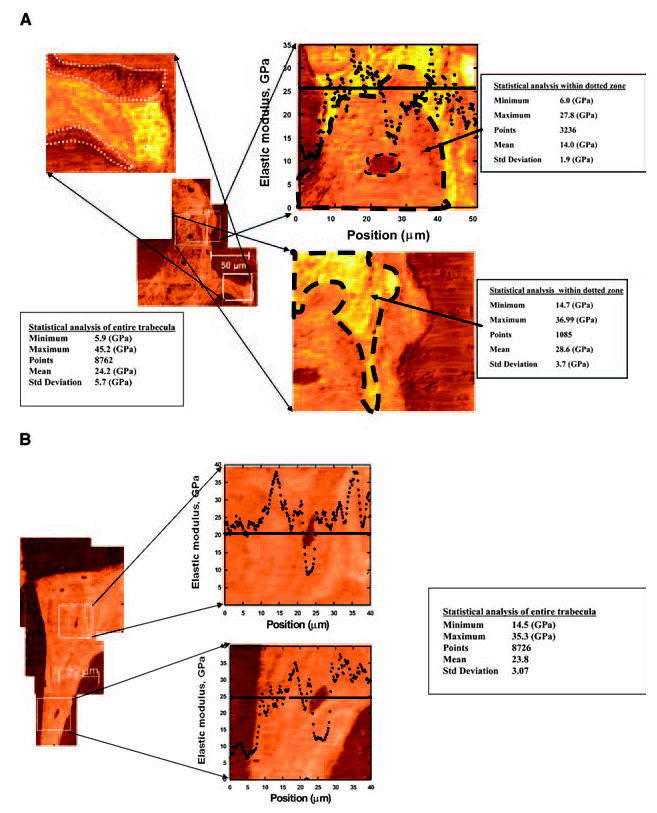
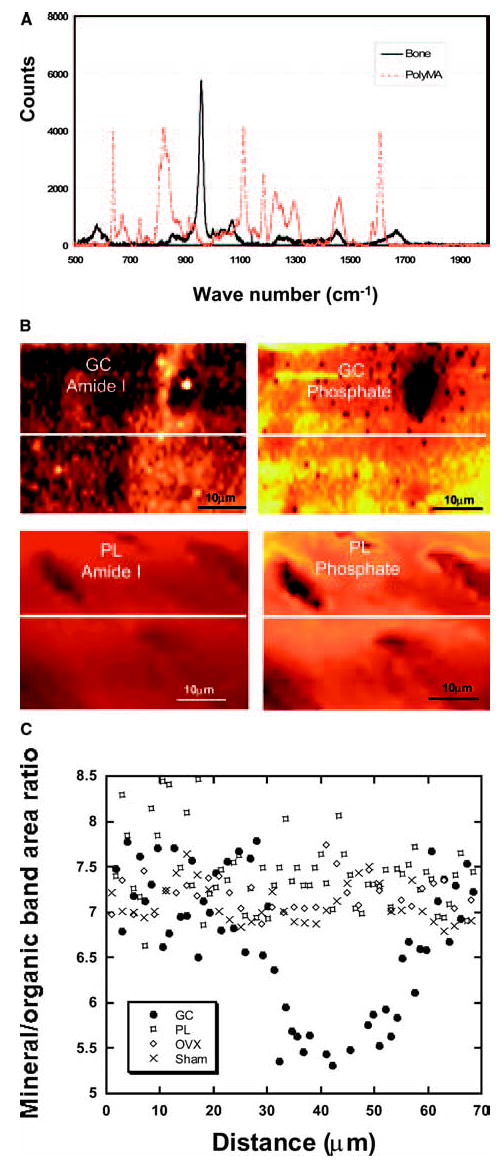
Results: Significant reductions in trabecular bone volume and whole bone strength occurred in both prednisolone-treated and estrogen-deficient mice compared with controls after 21 days (p < 0.05). The average elastic modulus over the entire surface of each trabecula was similar in all the experimental groups. However, localized changes within the trabeculae in areas surrounding the osteocyte lacunae were observed only in the prednisolone-treated mice. The size of the osteocyte lacunae was increased, reduced elastic modulus around the lacunae was observed, and a "halo" of hypomineralized bone surrounding the lacunae was observed. This was associated with reduced (nearly 40%) mineral to matrix ratio determined by Raman microspectroscopy. These localized changes in elastic modulus and bone mineral to matrix ratio were not observed in the other three experimental groups.
Conclusions: Based on these results, it seems that GCs may have direct effects on osteocytes, resulting in a modification of their microenvironment. These changes, including an enlargement of their lacunar space and the generation of a surrounding sphere of hypomineralized bone, seem to produce highly localized changes in bone material properties that may influence fracture risk.
Conflict of interest statement
The authors state that they have no conflicts of interest.
Figures

References
-
- Lane NE. An update on glucocorticoid-induced osteoporosis. Rheum Dis Clin North Am. 2001;27:235–254. - PubMed
-
- Saag KG. Glucocorticoid-induced osteoporosis. Endocrinol Metab Clin North Am. 2003;32:135–157. - PubMed
-
- Van Staa TP, Laan RF, Barton IP, Cohen S, Reid DM, Cooper C. Bone density threshold and other predictors of vertebral fractures in patients receiving oral glucocorticoid therapy. Arthritis Rheum. 2003;48:3224–3229. - PubMed
-
- Van Staa TP, Leufkens HS, Cooper C. The epidemiology of corticosteroid osteoporosis. A meta-analysis. Osteoporos Int. 2002;13:777–787. - PubMed
Publication types
MeSH terms
Substances
Grants and funding
LinkOut - more resources
Full Text Sources
Other Literature Sources
Medical
Miscellaneous
