

Comparison of biophysical and biologic properties of alpha-helical enantiomeric antimicrobial peptides
- PMID: 16492164
- PMCID: PMC3252236
- DOI: 10.1111/j.1747-0285.2006.00349.x
Comparison of biophysical and biologic properties of alpha-helical enantiomeric antimicrobial peptides
Abstract
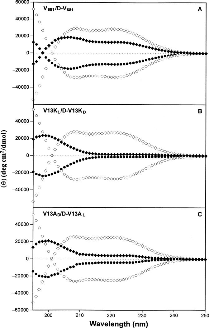
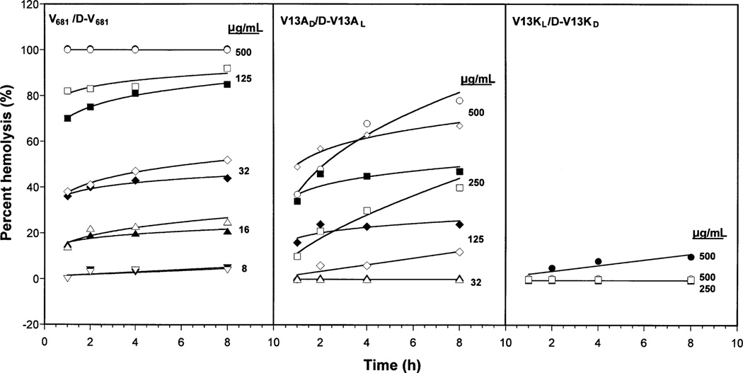
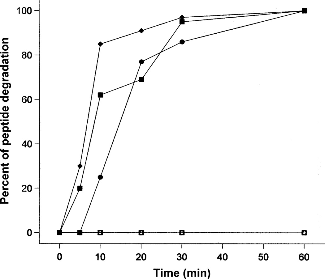
In our previous study (Chen et al. J Biol Chem 2005, 280:12316-12329), we utilized an alpha-helical antimicrobial peptide V(681) as the framework to study the effects of peptide hydrophobicity, amphipathicity, and helicity on biologic activities where we obtained several V(681) analogs with dramatic improvement in peptide therapeutic indices against gram-negative and gram-positive bacteria. In the present study, the D-enantiomers of three peptides--V(681), V13A(D) and V13K(L) were synthesized to compare biophysical and biologic properties with their enantiomeric isomers. Each D-enantiomer was shown by circular dichroism spectroscopy to be a mirror image of the corresponding L-isomer in benign conditions and in the presence of 50% trifluoroethanol. L- and D-enantiomers exhibited equivalent antimicrobial activities against a diverse group of Pseudomonas aeruginosa clinical isolates, various gram-negative and gram-positive bacteria and a fungus. In addition, L- and D-enantiomeric peptides were equally active in their ability to lyse human red blood cells. The similar activity of L- and D-enantiomeric peptides on prokaryotic or eukaryotic cell membranes suggests that there are no chiral receptors and the cell membrane is the sole target for these peptides. Peptide D-V13K(D) showed significant improvements in the therapeutic indices compared with the parent peptide V(681) by 53-fold against P. aeruginosa strains, 80-fold against gram-negative bacteria, 69-fold against gram-positive bacteria, and 33-fold against Candida albicans. The excellent stability of D-enantiomers to trypsin digestion (no proteolysis by trypsin) compared with the rapid breakdown of the L-enantiomers highlights the advantage of the D-enantiomers and their potential as clinical therapeutics.
Figures
Similar articles
-
Effects of net charge and the number of positively charged residues on the biological activity of amphipathic alpha-helical cationic antimicrobial peptides.Biopolymers. 2008;90(3):369-83. doi: 10.1002/bip.20911. Biopolymers. 2008. PMID: 18098173 Free PMC article.
-
Effects of single D-amino acid substitutions on disruption of beta-sheet structure and hydrophobicity in cyclic 14-residue antimicrobial peptide analogs related to gramicidin S.J Pept Res. 2004 Feb;63(2):69-84. doi: 10.1046/j.1399-3011.2003.00106.x. J Pept Res. 2004. PMID: 15009528 Free PMC article.
-
Antimicrobial and cytolytic properties of the frog skin peptide, kassinatuerin-1 and its L- and D-lysine-substituted derivatives.Peptides. 2005 Nov;26(11):2104-10. doi: 10.1016/j.peptides.2005.04.003. Peptides. 2005. PMID: 15885852
-
Fighting microbial infections: A lesson from amphibian skin-derived esculentin-1 peptides.Peptides. 2015 Sep;71:286-95. doi: 10.1016/j.peptides.2015.04.018. Epub 2015 May 8. Peptides. 2015. PMID: 25959536 Review.
-
Advances in antimicrobial peptide immunobiology.Biopolymers. 2006;84(5):435-58. doi: 10.1002/bip.20543. Biopolymers. 2006. PMID: 16736494 Review.
Cited by
-
In silico design and optimization of selective membranolytic anticancer peptides.Sci Rep. 2019 Aug 2;9(1):11282. doi: 10.1038/s41598-019-47568-9. Sci Rep. 2019. PMID: 31375699 Free PMC article.
-
In vitro activities of synthetic host defense propeptides processed by neutrophil elastase against cystic fibrosis pathogens.Antimicrob Agents Chemother. 2011 May;55(5):2487-9. doi: 10.1128/AAC.01384-10. Epub 2011 Feb 22. Antimicrob Agents Chemother. 2011. PMID: 21343449 Free PMC article.
-
HPLC analysis and purification of peptides.Methods Mol Biol. 2007;386:3-55. doi: 10.1007/978-1-59745-430-8_1. Methods Mol Biol. 2007. PMID: 18604941 Free PMC article.
-
Effect of BMAP-28 antimicrobial peptides on Leishmania major promastigote and amastigote growth: role of leishmanolysin in parasite survival.PLoS Negl Trop Dis. 2011;5(5):e1141. doi: 10.1371/journal.pntd.0001141. Epub 2011 May 31. PLoS Negl Trop Dis. 2011. PMID: 21655347 Free PMC article.
-
Preparative reversed-phase high-performance liquid chromatography collection efficiency for an antimicrobial peptide on columns of varying diameters (1mm to 9.4mm I.D.).J Chromatogr A. 2007 Jan 26;1140(1-2):112-20. doi: 10.1016/j.chroma.2006.11.052. Epub 2006 Dec 6. J Chromatogr A. 2007. PMID: 17156789 Free PMC article.
References
-
- Travis J. Reviving the antibiotic miracle? Science. 1994;264:360–362. - PubMed
-
- Neu HC. The crisis in antibiotic resistance. Science. 1992;257:1064–1073. - PubMed
-
- Hancock RE. Peptide antibiotics. Lancet. 1997;349:418–422. - PubMed
-
- Andreu D, Rivas L. Animal antimicrobial peptides: an overview. Biopolymers. 1998;47:415–433. - PubMed
-
- Sitaram N, Nagaraj R. Host-defense antimicrobial peptides: importance of structure for activity. Curr Pharm Des. 2002;8:727–742. - PubMed
Publication types
MeSH terms
Substances
Grants and funding
LinkOut - more resources
Full Text Sources
Other Literature Sources
