

Review
doi: 10.1186/jbiol31.
Epub 2005 Jan 9.
Recycling signals in the neural crest
Affiliations
- PMID: 16492316
- PMCID: PMC1414065
- DOI: 10.1186/jbiol31
Item in Clipboard
Review
Recycling signals in the neural crest
J Biol.
2005.
Abstract
Vertebrate neural crest cells are multipotent and differentiate into structures that include cartilage and the bones of the face, as well as much of the peripheral nervous system. Understanding how different model vertebrates utilize signaling pathways reiteratively during various stages of neural crest formation and differentiation lends insight into human disorders associated with the neural crest.
Figures
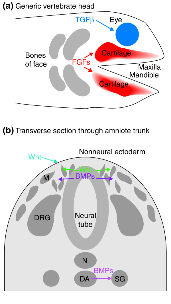
Recycling counts in the neural crest. The reiterative function of various signaling molecules (Wnts, TGFβ/BMPs, and FGFs) is tantamount to the regulation of neural crest development at multiple stages, ranging from the initial phases of induction to migration and subsequent differentiation. Depending upon their developmental stage, neural crest cells respond differently to the same signals. (a) Neural crest cells build much of the facial skeleton. TGFβ and FGF molecules signal to ensure proper development of the eye and facial cartilage, respectively. (b) In the trunk, Wnts and BMPs work to specify various neural crest derivatives. Early Wnt signals from the nonneural ectoderm are important in neural crest induction, whereas later Wnts specify neural crest cells to become sensory neurons and pigment cells. In addition, BMPs, also members of the TGFβ family, are produced by the dorsal aorta to regulate sympathetic neuron differentiation. DA, dorsal aorta; DRG, dorsal root ganglion; SG, sympathetic ganglion; N, notochord; M, melanocyte.
References
Publication types
MeSH terms
Grants and funding
LinkOut - more resources
Full Text Sources
