. 2006 Feb 28;103(9):3250-5.
doi: 10.1073/pnas.0600012103. Epub 2006 Feb 21.

Progressive vascular smooth muscle cell defects in a mouse model of Hutchinson-Gilford progeria syndrome

Affiliations

Progressive vascular smooth muscle cell defects in a mouse model of Hutchinson-Gilford progeria syndrome

Renee Varga et al. Proc Natl Acad Sci U S A. .

Abstract

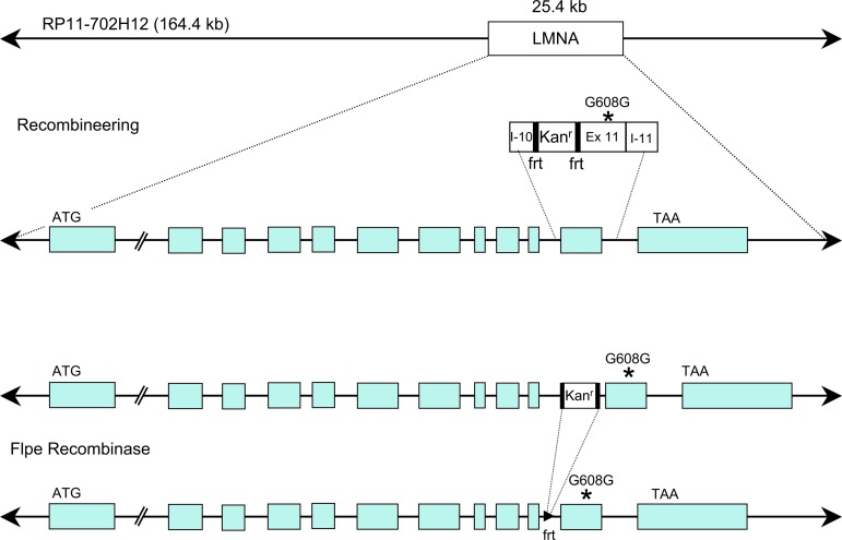
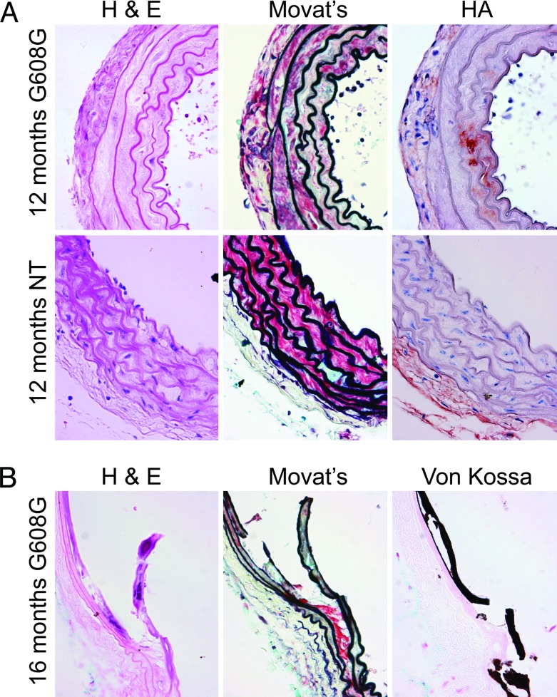
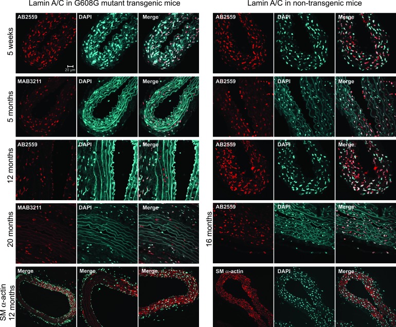
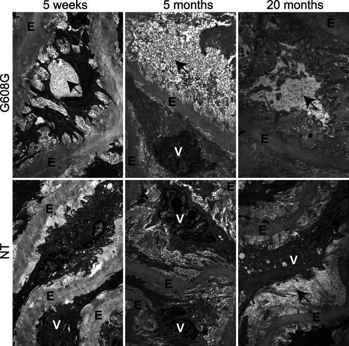
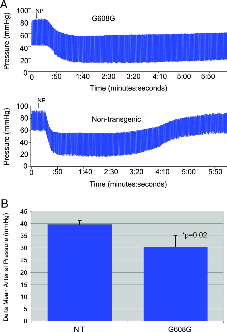
Children with Hutchinson-Gilford progeria syndrome (HGPS) suffer from dramatic acceleration of some symptoms associated with normal aging, most notably cardiovascular disease that eventually leads to death from myocardial infarction and/or stroke usually in their second decade of life. For the vast majority of cases, a de novo point mutation in the lamin A (LMNA) gene is the cause of HGPS. This missense mutation creates a cryptic splice donor site that produces a mutant lamin A protein, termed "progerin," which carries a 50-aa deletion near its C terminus. We have created a mouse model for progeria by generating transgenics carrying a human bacterial artificial chromosome that harbors the common HGPS mutation. These mice develop progressive loss of vascular smooth muscle cells in the medial layer of large arteries, in a pattern very similar to that seen in children with HGPS. This mouse model should prove valuable for testing experimental therapies for this devastating disorder and for exploring cardiovascular disease in general.


Conflict of interest statement

Conflict of interest statement: No conflicts declared.

Figures

Fig. 1.
Fig. 1.
Procedure for generating the BAC G608G transgenic construct by homologous recombination in E. coli. Human BAC clone RP11-702H12 contains an insert of 164.4 kb of genomic DNA from chromosome 1q including the LMNA gene. Homologous recombination was induced between this BAC and a shuttle fragment of 2.3 kb containing the common G608G HGPS mutation within exon 11 (Ex 11) of LMNA and the kanamycin resistance gene (Kanr) flanked by two FRT sites. Homology arms with sequences from intron 10 (I-10) and intron 11 (I-11) of the LMNA gene were used to direct the recombination event between the shuttle fragment and the BAC clone. After selection for positive recombinants, Flpe recombinase was induced to release the kanamycin gene. The final BAC G608G transgenic construct used for pronuclear injection differs from the original BAC in that it contains the G608G mutation (∗) within exon 11 of LMNA and 109 extra nucleotides in intron 10 including an FRT site, EcoRI site, and SacI site.
Fig. 2.
Fig. 2.
Progressive arterial abnormalities in progeria mice. (A) Analysis of aortic sections of 12-month-old mice by using hematoxylin/eosin (H & E) and Movat's pentachrome stain and immunohistochemistry analysis for hyaluronan indicates a progressive loss of VSMC, elastin breakage, thickened medial layer and adventitia, and PG accumulation. (B) All G608G mice examined over 16 months of age showed evidence of carotid (shown) and aortic (data not shown) calcification, which was confirmed with Von Kossa staining. Younger HGPS and all ages of NT mice showed no evidence of calcification. HA, hyaluronan.
Fig. 3.
Fig. 3.
Lamin A immunofluorescent staining of aortic sections highlights the progressive VSMC loss in G608G transgenics. The presence of lamin A/C is detected in descending aorta by using either the mAb3211 antibody (detects human but not mouse) or the Ab2559 antibody (detects both human and mouse). The age of each animal is indicated on the figure. The nuclei are counterstained with DAPI (blue), and an overlay of the lamin A/C and DAPI images is presented (merge). Cells with lamin A/C staining are present in the three layers of the arteries: intima, media, and adventitia in 5-week-old and 5-month-old transgenic mice. The number of lamin A/C-positive nuclei is dramatically decreased in the 12-month-old mice. They are almost completely absent in the medial layer of older transgenic animals (20 months). Note the presence of “ghost” nuclei in the 12- and 20-month-old G608G mice that retain lamin A/C staining, but lack DAPI staining. Smooth muscle α-actin staining confirms loss of VSMCs; these images also show the eccentric distribution of vascular damage, with some areas around the vessel circumference better preserved than others.
Fig. 4.
Fig. 4.
Transmission electron microscopy comparing nontransgenic (NT) and G608G H-line carotid artery sections showing in the G608G line the progressive accumulation of cellular debris, frayed elastin fibers, and accumulation and disorganization of collagen and PG (see black arrows). The nontransgenic vessel shows only mild changes at 20 months. E, elastin; V, VSMC.
Fig. 5.
Fig. 5.
Diminished vascular responsiveness in progeria mice. (A) Representative blood pressure tracings taken before and after NP administration in 5-month-old mice. (B) The drop in mean arterial pressure, comparing blood pressure before and after the administration of NP, is blunted in the G608G mice as compared with the control mice at 5 months of age.

Similar articles

Cited by

References

    1. DeBusk F. L. J. Pediatr. (Berlin) 1972;80:697–724. - PubMed
    1. Baker P. B., Baba N., Boesel C. P. Arch. Pathol. Lab. Med. 1981;105:384–386. - PubMed
    1. Stehbens W. E., Wakefield S. J., Gilbert-Barness E., Olson R. E., Ackerman J. Cardiovasc. Pathol. 1999;8:29–39. - PubMed
    1. Stehbens W. E., Delahunt B., Shozawa T., Gilbert-Barness E. Cardiovasc. Pathol. 2001;10:133–136. - PubMed
    1. Ackerman J., Gilbert-Barness E. Pediatr. Pathol. Mol. Med. 2002;21:1–13. - PubMed

Publication types

MeSH terms

LinkOut - more resources