. 2006 Feb 28;103(9):3416-21.
doi: 10.1073/pnas.0511290103. Epub 2006 Feb 21.

Prion protein (PrPc) positively regulates neural precursor proliferation during developmental and adult mammalian neurogenesis

Affiliations

Prion protein (PrPc) positively regulates neural precursor proliferation during developmental and adult mammalian neurogenesis

Andrew D Steele et al. Proc Natl Acad Sci U S A. .

Abstract

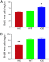
The misfolding of the prion protein (PrP(c)) is a central event in prion diseases, yet the normal function of PrP(c) remains unknown. PrP(c) has putative roles in many cellular processes including signaling, survival, adhesion, and differentiation. Given the abundance of PrP(c) in the developing and mature mammalian CNS, we investigated the role of PrP(c) in neural development and in adult neurogenesis, which occurs constitutively in the dentate gyrus (DG) of the hippocampus and in the olfactory bulb from precursors in the subventricular zone (SVZ)/rostral migratory stream. In vivo, we find that PrP(c) is expressed immediately adjacent to the proliferative region of the SVZ but not in mitotic cells. In vivo and in vitro studies further find that PrP(c) is expressed in multipotent neural precursors and mature neurons but is not detectable in glia. Loss- and gain-of-function experiments demonstrate that PrP(c) levels correlate with differentiation of multipotent neural precursors into mature neurons in vitro and that PrP(c) levels positively influence neuronal differentiation in a dose-dependent manner. PrP(c) also increases cellular proliferation in vivo; in the SVZ, PrP(c) overexpresser (OE) mice have more proliferating cells compared with wild-type (WT) or knockout (KO) mice; in the DG, PrP(c) OE and WT mice have more proliferating cells compared with KO mice. Our results demonstrate that PrP(c) plays an important role in neurogenesis and differentiation. Because the final number of neurons produced in the DG is unchanged by PrP(c) expression, other factors must control the ultimate fate of new neurons.


Conflict of interest statement

Conflict of interest statement: No conflicts declared.

Figures

Fig. 1.
Fig. 1.
Confirmation of PrP protein expression and in vivo antibody specificity. (A) The absence of PrPc in KO mouse brain and its overexpression in PrPc OE transgenic mice was confirmed by Western blot analysis of whole-brain homogenate by using a monoclonal antibody against the PrPc protein. The top, middle, and lower bands of the PrPc blot (Upper) correspond to di-, mono-, and unglycosylated PrPc, respectively. The blot was reprobed with an antibody against TuJ1 to demonstrate equal loading (Lower). (B and C) Specificity of the polyclonal antibody against PrPc was confirmed in 30-μm sections of adult brain from PrPc OE (B) and PrPc KO (C) mice at equal exposures. cc, corpus callosum; ctx, cortex; hpc, hippocampus.
Fig. 2.
Fig. 2.
PrPc is expressed in neurogenic regions and is strongly expressed in neurons in vivo and in vitro. (A) Proliferation in the SVZ of the adult mouse detected with BrdUrd (green, arrowheads). PrPc-positive cells (red, arrows) are found adjacent to the neurogenic region. (B) A close-up of the SVZ region shown in (A), indicating that PrPc (red, arrows) is not expressed in proliferating cells (green, arrowheads) but instead is expressed in cells just lateral to those proliferating in the SVZ. (C and D) 3D confocal reconstructions of the CNS in vivo. (C) PrPc (red in all micrographs) is strongly expressed in mature NeuN-positive neurons (green, arrows). Note the lack of PrPc in surrounding DAPI-stained cells with small compact nuclei consistent with glia (arrowheads). (D) PrPc is not expressed in GFAP-positive astroglia identified by a human GFAP (hGFAP) promoter driving expression of eGFP (arrowhead, green). PrPc-positive GFAP-negative cells (arrows) surround the astrocyte. (E and F) Embryonic neural precursor cultures. (E) PrPc (red, arrows) is most strongly expressed in MAP-2-positive neurons (green), whereas it is not detected in S100β-positive astroglia (F) (arrowheads, green). DAPI nuclear counterstain (blue) in B, C, E, and F. cc, corpus callosum; lv, lateral ventricle. (Scale bars: A, 100 μm; B, 10 μm; C and D, 25 μm; E and F, 100 μm.)
Fig. 3.
Fig. 3.
PrPc levels increase differentiation of embryonic neural precursors in vitro. (A) Neural precursors derived from PrPc KO embryos remain as uncommitted multipotent precursors (A′; nestin-positive, arrow) for a longer period than do precursor derived from WT and, especially, OE embryos. Three days after induction of precursor differentiation via removal of basic fibroblast growth factor (3 DIV), there are significant differences between the percentage of nestin-positive precursors derived from KO, WT, and OE embryos. PrPc OE precursors differentiate from their multipotent state more rapidly than do those from KO and WT mice, even at 1 DIV. By 7 DIV, there are no significant differences among any of the three groups. (B) Differentiation and maturation into a neuronal phenotype (B′; MAP-2-positive; cell body, arrow; cell process, arrowhead) occurs at a significantly slower rate in PrPc KO-derived precursors than in WT, and differentiation is significantly more rapid in PrPc OE-derived precursors. PrPc KO precursors are still capable of producing neurons, indicating a delay in neuronal production rather than a failure to differentiate. ∗, P < 0.05; ∗∗, P < 0.01; ∗∗∗, P < 0.001. (Scale bars: A′ and B′, 50 μm.)
Fig. 4.
Fig. 4.
PrPc increases proliferation in vivo. (A) One hour after a pulse label of BrdUrd, PrPc OE mice have significantly more proliferating cells in the SVZ than in PrPc KO or WT mice. (B) In the DG, PrPc KO mice have significantly fewer proliferating cells than do WT or OE mice. ∗, P < 0.01; ∗∗, P < 0.001.

References

    1. Prusiner S. B. Proc. Natl. Acad. Sci. USA. 1998;95:13363–13383. - PMC - PubMed
    1. Aguzzi A., Polymenidou M. Cell. 2004;116:313–327. - PubMed
    1. Hetz C., Maundrell K., Soto C. Trends Mol. Med. 2003;9:237–243. - PubMed
    1. Bueler H., Aguzzi A., Sailer A., Greiner R. A., Autenried P., Aguet M., Weissmann C. Cell. 1993;73:1339–1347. - PubMed
    1. Kanaani J., Prusiner S. B., Diacovo J., Baekkeskov S., Legname G. J. Neurochem. 2005;95:1373–1386. - PubMed

Publication types