. 2006 Feb 28;103(9):3298-303.
doi: 10.1073/pnas.0511233103. Epub 2006 Feb 21.

IL-32, a proinflammatory cytokine in rheumatoid arthritis

Affiliations

IL-32, a proinflammatory cytokine in rheumatoid arthritis

Leo A B Joosten et al. Proc Natl Acad Sci U S A. .

Abstract

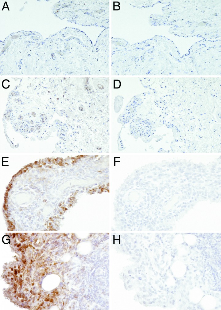
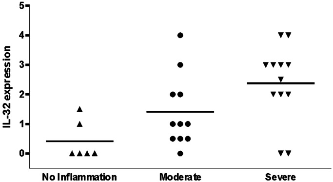
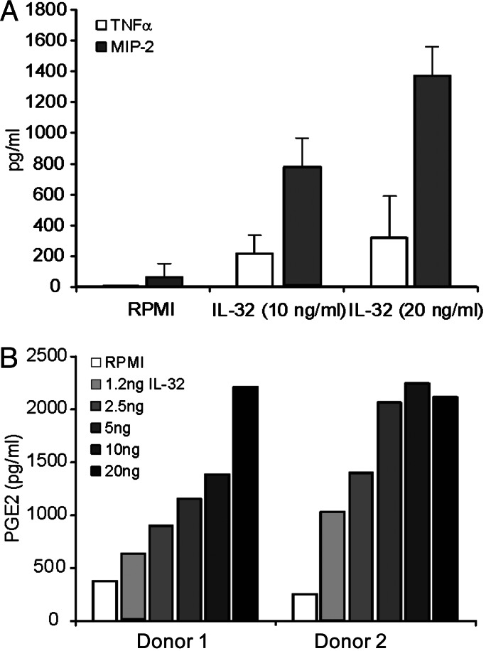
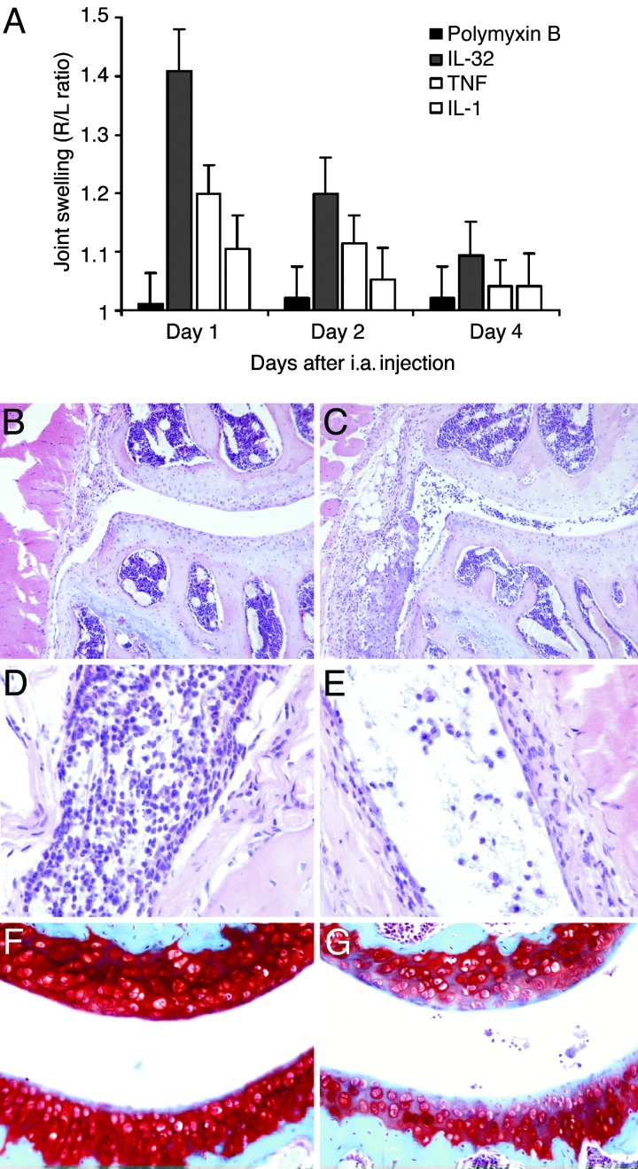
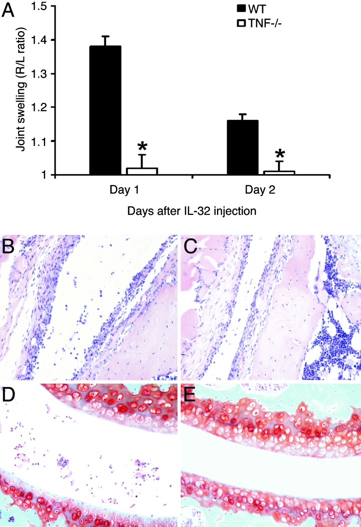
IL-32 is a recently discovered cytokine that induces TNFalpha, IL-1beta, IL-6, and chemokines. We investigated whether IL-32 is expressed in the synovia of patients with rheumatoid arthritis (RA) and studied associations with disease severity and the presence of other cytokines. Immunohistochemistry revealed that IL-32 is highly expressed in RA synovial tissue biopsies, whereas IL-32 was not observed in synovial tissues from patients with osteoarthritis. Moreover, in synovial biopsies from 29 RA patients with active disease, the level of IL-32 staining correlated with erythrocyte sedimentation rate, a marker of systemic inflammation (R = 0.63 and P < 0.0003). Synovial staining of IL-32 also correlated with indices of synovial inflammation (R = 0.80 and P < 0.0001) as well as synovial presence of TNFalpha (R = 0.68 and P < 0.004), IL-1beta (R = 0.79 and P < 0.0001), and IL-18 (R = 0.82 and P < 0.001). IL-32 was a potent inducer of prostaglandin E(2) release in mouse macrophages and human blood monocytes, an important property for inflammation. After the injection of human IL-32gamma into the knee joints of naïve mice, joint swelling, with pronounced influx of inflammatory cells and cartilage damage, was observed. In TNFalpha-deficient mice, IL-32-driven joint swelling was absent and cell influx was markedly reduced, but loss of proteoglycan was unaffected, suggesting that IL-32 activity is, in part, TNFalpha-dependent. IL-32, strongly associated with TNFalpha, IL-1beta, and IL-18, appears to play a role in human RA and may be a novel target in autoimmune diseases.


Conflict of interest statement

Conflict of interest statement: No conflicts declared.

Figures

Fig. 1.
Fig. 1.
IL-32 expression in RA synovial tissue biopsies. (A) Synovial tissue sample from a healthy individual. (C) RA synovial tissue, not inflamed. (E) RA synovial tissue, moderately inflamed. (G) RA synovial tissue, severely inflamed. (B, D, F, and H) Control staining was performed with nonrelevant isotope control antibody. H&E staining was performed as counterstaining. (Original magnification ×400.)
Fig. 2.
Fig. 2.
Expression of synovial IL-32 in relation to macroscopic knee scores of patients with RA. On macroscopic scoring, the knee joints that underwent percutaneous biopsing were categorized as noninflamed, moderately inflamed, and severely inflamed. Staining for IL-32 was semiquantitatively scored on a five-point scale (range 0–4) at ×200 magnification; a score of 0 represented no or minimal staining, a score of 1 indicated 10–20% positive cells, a score of 2 indicated 30–40% positive cells, a score of 3 indicated 50–60% positive cells, and a score of 4 represented staining of >60% of the cells. Data were derived from 29 RA patients with active disease.
Fig. 3.
Fig. 3.
Production of proinflammatory mediators by IL-32. (A) TNFα and MIP-2 production of peritoneal macrophages isolated from LPS-resistant mice (C3H/HeJ mice). Murine TNFα and MIP-2 were measured after 24-h IL-32 exposure by electrochemiluminescence (13). Data are the mean ± SD levels of three identical experiments. (B) PGE2 production of human PBMC stimulated with IL-32. PBMC isolated from two healthy donors were exposed for 24 h to IL-32 in a dose ranging from 1.25 ng/ml to 20 ng/ml. PGE2 concentrations were measured by enhanced electrochemiluminescence with acetylcholinesterase-conjugated tracer used for quantification. The sensitivity of the assay was 25 pg/ml.
Fig. 4.
Fig. 4.
Joint inflammation provoked by local IL-32γ injection. (A) Joint swelling after i.a. injection of 100 ng of recombinant IL-32γ into the right knee joints of C57/Bl6 mice, determined by the 99mTc-uptake method. IL-32 was compared with 100 ng of either murine TNFα or murine IL-1β. Polymyxin B (7 ng) was injected as control. (B) Histopathology at day 2 after i.a. injection of polymyxin B (7 ng). H&E staining was performed. (Original magnification, ×100.) (C) Joint inflammation after i.a. injection of 100 ng of IL-32γ. H&E staining was performed. (Original magnification, ×100.) (D) Severe cell influx in joint cavity and synovial tissue. H&E staining was performed. (Original magnification, ×200.) (E) Monocyte/macrophage-like cells at day 4 after IL-32γ injection. H&E staining was performed. (Original magnification, ×400.) (F) No cartilage matrix proteoglycan loss at day 4 after polymyxin B injection, visualized by Safranin O staining. (Original magnification, ×200.) (G) Depletion of cartilage proteoglycans at day 4 after IL-32γ exposure.
Fig. 5.
Fig. 5.
IL-32-driven joint inflammation in TNFα-deficient mice. (A) Joint swelling was determined at days 1 and 2 after i.a. injection of 100 ng of IL-32γ into WT or TNFα-deficient (TNF−/−) mice. As control, 6 μl of polymyxin B (7 ng) was injected in the left knee joint (data not shown). The data represent five mice per group. ∗, P < 0.001, Mann–Whitney U test, compared with WT mice. (B) Synovitis at day 2 after i.a. injection of 100 ng of IL-32γ into the knee of a WT mouse. H&E staining was performed. (Original magnification, ×200.) (C) Cell influx in the synovial lining of same dose of IL-32 in TNFα-deficient mice. (D and E) Proteoglycan loss in patella and femur cartilage layers in both WT and TNFα-deficient mice. Safranin O staining was performed. (Original magnification, ×200.) See Fig. 4F for normal cartilage proteoglycan staining.

Similar articles

Cited by

References

    1. Arend W. P., Dayer J.-M. Arthritis Rheum. 1995;38:151–160. - PubMed
    1. Feldmann M., Brennan F. M., Maini R. N. Annu. Rev. Immunol. 1996;14:397–440. - PubMed
    1. Miossec P., van den Berg W. Arthritis Rheum. 1997;40:2105–2115. - PubMed
    1. Bresnihan B., Tak P. P. Baillieres Best. Pract. Res. Clin. Rheumatol. 1999;13:645–659. - PubMed
    1. Goronzy J. J., Weyand C. M. Immunol. Rev. 2005;204:55–73. - PubMed

Publication types