. 2006 Feb 28;103(9):3072-7.
doi: 10.1073/pnas.0510580103. Epub 2006 Feb 21.

Structure-based inhibitor design of AccD5, an essential acyl-CoA carboxylase carboxyltransferase domain of Mycobacterium tuberculosis

Affiliations

Structure-based inhibitor design of AccD5, an essential acyl-CoA carboxylase carboxyltransferase domain of Mycobacterium tuberculosis

Ting-Wan Lin et al. Proc Natl Acad Sci U S A. .

Abstract

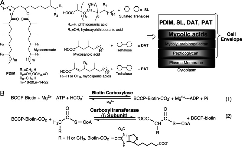
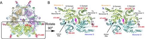
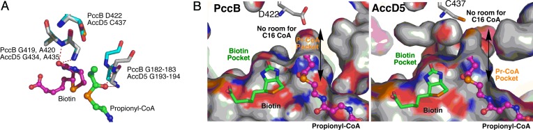
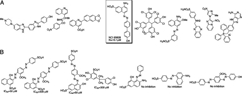
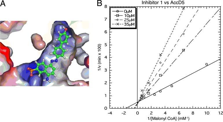
Mycolic acids and multimethyl-branched fatty acids are found uniquely in the cell envelope of pathogenic mycobacteria. These unusually long fatty acids are essential for the survival, virulence, and antibiotic resistance of Mycobacterium tuberculosis. Acyl-CoA carboxylases (ACCases) commit acyl-CoAs to the biosynthesis of these unique fatty acids. Unlike other organisms such as Escherichia coli or humans that have only one or two ACCases, M. tuberculosis contains six ACCase carboxyltransferase domains, AccD1-6, whose specific roles in the pathogen are not well defined. Previous studies indicate that AccD4, AccD5, and AccD6 are important for cell envelope lipid biosynthesis and that its disruption leads to pathogen death. We have determined the 2.9-Angstroms crystal structure of AccD5, whose sequence, structure, and active site are highly conserved with respect to the carboxyltransferase domain of the Streptomyces coelicolor propionyl-CoA carboxylase. Contrary to the previous proposal that AccD4-5 accept long-chain acyl-CoAs as their substrates, both crystal structure and kinetic assay indicate that AccD5 prefers propionyl-CoA as its substrate and produces methylmalonyl-CoA, the substrate for the biosyntheses of multimethyl-branched fatty acids such as mycocerosic, phthioceranic, hydroxyphthioceranic, mycosanoic, and mycolipenic acids. Extensive in silico screening of National Cancer Institute compounds and the University of California, Irvine, ChemDB database resulted in the identification of one inhibitor with a K(i) of 13.1 microM. Our results pave the way toward understanding the biological roles of key ACCases that commit acyl-CoAs to the biosynthesis of cell envelope fatty acids, in addition to providing a target for structure-based development of antituberculosis therapeutics.


Conflict of interest statement

Conflict of interest statement: No conflicts declared.

Figures

Fig. 1.
Fig. 1.
Chemical structures of cell wall fatty acids and ACCase mechanism. (A) The chemical structures of mycolic acids and multimethyl-branched fatty acids such as mycocerosic, phthioceranic, hydroxyphthioceranic, mycosanoic, and mycolipenic acids, which form liposugars phthiocerol dimycocerosate (PDIM), sulfated tetraacyl trehalose (SL1), diacyl trehaloses (DAT1), triacyl trehalose (TAT), and pentaacyl trehalose (PAT), respectively. A proposed cell envelope architecture is shown, adopted from ref. . (B) Acyl-CoA carboxylase (ACCase) provides the extender units for the biosyntheses of cell envelope fatty acids. The α [biotin carboxylase (BC) and biotin carboxylate carrier protein (BCCP)] and β [carboxyltransferase (CT)] subunits catalyze the first and second steps, respectively. The β subunit is the key domain that determines the ACCase substrate specificity. In M. tuberculosis, there are six β subunits, AccD1–6, whose biological roles are not well defined.
Fig. 2.
Fig. 2.
The AccD5 crystal structure. (A) Overall structure of AccD5 as two stacks of trimers. Only minor differences are observed among the six monomers, with an rms deviation of <0.4 Å. The threefold axis of monomers A–B–C and D–E–F is indicated. The loops in red are regions that are different between AccD5 and PccB. The N-domain helices (α1, α2, and α3) of monomer A, B, or C interact extensively with the C-domain helices (α18, α19, and α20) of monomer D, E, or F. (B) Stereoview of the dimeric, di-domain interactions, shown between monomers A (in yellow) and D (in blue) are important for protein stability, enzyme catalysis, and substrate specificity.
Fig. 3.
Fig. 3.
The AccD5 active site. (A) Structural overlap between AccD5 and PccB near the active site. Two sets of oxyanion-stabilizing residues are highly conserved, including the NH of G193 and G194 and the NH of G434′ and A435′. The structural similarity strongly suggests a similar enzyme mechanism. (B) Although surfaces outside of the pocket are quite different, the size and shape of the acyl-CoA-binding pockets themselves are very similar between PccB and AccD5, whose pockets are too small to accommodate the 16-carbon palmitoyl-CoA.
Fig. 4.
Fig. 4.
The in silico inhibitor leads of AccD5. (A) The lead compounds from the first round of in silico inhibitor screening against the AccD5 active site, in which only NCI-65828 showed extensive enzyme inhibition. (B) The lead compounds from the second round of in silico screening of 3,000 chemical homologs that resemble NCI-65828. The IC50 values of these analogs range from 25 to 300 μM, with >50% lacking inhibitory effect on AccD5.
Fig. 5.
Fig. 5.
After extensive in silico screening, the ligand NCI-65828 was found to inhibit AccD5 competitively with an experimental Ki of 13.1 μM. (A) Docking of NCI-65828 in the acyl-CoA-binding pocket of AccD5 matches the binding motif of an acyl-CoA, in which the anionic sulfate of NCI-65828 binds the entrance of the CoA pocket and the hydrophobic moiety binds the hydrophobic interior of the CoA pocket. (B) The Lineweaver–Burk plot shows that NCI-65828 is a competitive inhibitor of AccD5.

References

    1. Ishikawa N. Kekkaku. 2005;80:89–94. - PubMed
    1. Kunimoto D., Long R. Respir. Care Clin. N. Am. 2005;11:25–34. - PubMed
    1. Bates I., Fenton C., Gruber J., Lalloo D., Lara A. M., Squire S. B., Theobald S., Thomson R., Tolhurst R. Lancet Infect. Dis. 2004;4:368–375. - PubMed
    1. Bates I., Fenton C., Gruber J., Lalloo D., Medina Lara A., Squire S. B., Theobald S., Thomson R., Tolhurst R. Lancet Infect. Dis. 2004;4:267–277. - PubMed
    1. Chopra K. Indian J. Pediatr. 1996;63:159–162. - PubMed

Publication types

MeSH terms

Associated data

LinkOut - more resources