. 2006 Feb 28;103(9):3238-43.
doi: 10.1073/pnas.0509650103. Epub 2006 Feb 21.

High frequency of centromere inactivation resulting in stable dicentric chromosomes of maize

Affiliations

High frequency of centromere inactivation resulting in stable dicentric chromosomes of maize

Fangpu Han et al. Proc Natl Acad Sci U S A. .

Abstract

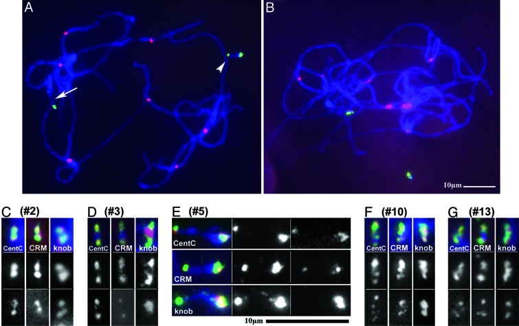
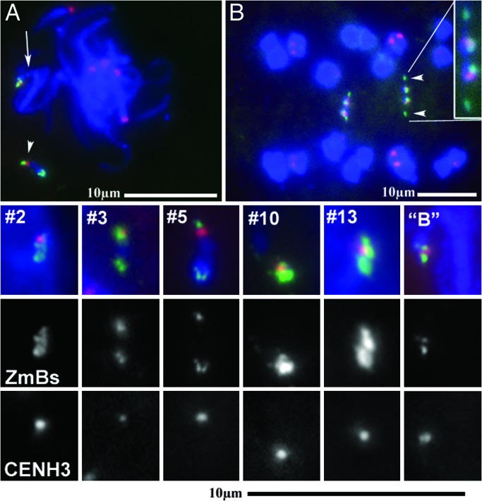
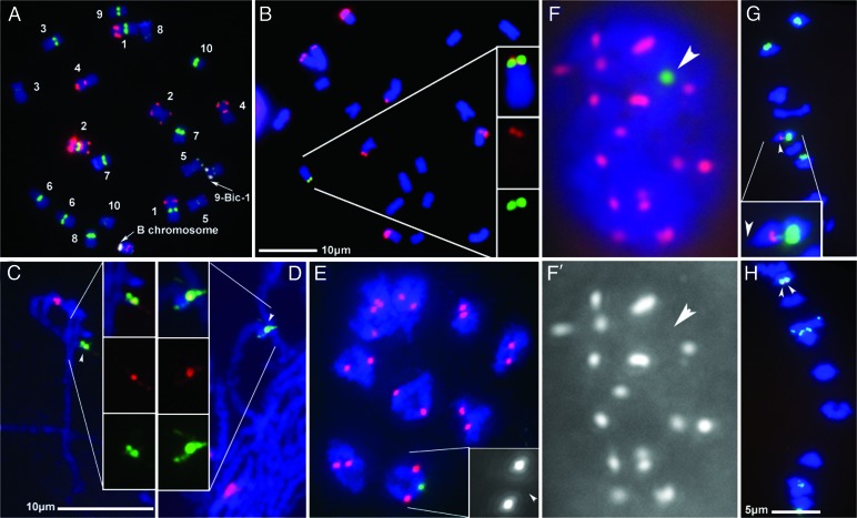
Somatic chromosome spreads from maize (Zea mays L.) plants containing B-A translocation chromosomes undergoing the chromosome type breakage-fusion-bridge cycle were examined by FISH. The size and type of extra chromosomes varied among cells of the same individual. A collection of minichromosomes derived from the chromosome type breakage-fusion-bridge cycle was examined for the presence of stable dicentric chromosomes. Six of 23 chromosomes in the collection contained two regions with DNA sequences typical of centromeres. Functional analysis and immunolabeling of CENH3, the centromere-specific histone H3 variant, revealed only one functional centromere per chromosome, despite the duplicate centromere sequences. One plant was found with an inactive B centromere that had been translocated to the short arm of chromosome 9. The translocated centromere region appeared identical to that of a normal B chromosome. The inactivation of the centromeres was stable for at least four generations. By using dicentrics from dispensable chromosomes, centromere inactivation was found to be quite common under these circumstances.


Conflict of interest statement

Conflict of interest statement: No conflicts declared.

Figures

Fig. 1.
Fig. 1.
The B9-Dp9 chromosome initiates the BFB cycle after crossing over with itself. (A) The duplicated portion of B9-Dp9 pairs with itself during meiosis I, and a crossover creates a dicentric chromosome and an acentric fragment. (B) During meiosis II, the sister centromeres migrate to opposite poles, and the resulting chromatin bridge is broken. (C) A chromosome with a broken end is delivered to the microsporocyte. (D) In the microsporocyte, the broken chromosome is replicated and fuses with itself. The centromeres migrate to opposite poles during the first pollen mitotic division, breaking the chromosome. (E) After the first pollen division, the broken chromosome is replicated in the generative cell. Because the B centromere usually undergoes nondisjunction during the second pollen division, both centromeres move to one pole. (F) In the subsequent somatic division after fertilization, the dicentric chromosome is replicated, and the centromeres could move to the poles independently of one another. If two centromeres on the same chromatid move to opposite poles, a double bridge is formed that might break at different locations. The broken ends of the chromosomes in each daughter cell can fuse, leading to another round of the chromosome type BFB cycle. As this process continues, chromosomes can be stabilized by inactivation of one of the centromeres or by healing of a broken end by addition of telomeres, leading to a variety of minichromosomes.
Fig. 2.
Fig. 2.
BFB cycle in root tips. A plant containing one B9-Dp9 and two 9-B translocation chromosomes was crossed as a male to a yg2, bz1 tester line. The resulting kernels were germinated, chromosome spreads prepared from the root tips, and FISH performed by using the B chromosome-specific ZmBs probe (green) and the 180-bp knob heterochromatin probe (red). Lack of crossing over in the duplicated region, followed by nondisjunction during the second pollen mitosis, results in two intact B9-Dp9 chromosomes as in A. (BD) Different cells from a single root tip vary in the size and number of extra chromosomes. The arrowhead indicates the ZmBs signal at the tip of the 9-B chromosome, and arrows indicate the ZmBs signal of the centromeric region.
Fig. 3.
Fig. 3.
Pachytene FISH of dicentric minichromosomes. (A) A pachytene spread with an intact B chromosome (indicated by the arrow) and minichromosome 5 hybridized with ZmBs (green) and CRM (red). The arrowhead indicates the active centromere of minichromosome 5. (B) A pachytene spread with two unpaired copies of minichromosome 10 hybridized with ZmBs (green) and CRM (red) illustrates the relative size of the minichromosomes. (CG) Minichromosomes 2, 3, 5, 10, and 13, respectively, are depicted from pachytene spreads. Chromosomes were hybridized with ZmBs (green) and CentC, CRM, or the 180-bp knob repeat (red). The gray values for the probes are also displayed, first ZmBs and, second, the indicated repetitive probe (CentC, CRM, or the 180-bp knob repeat).
Fig. 4.
Fig. 4.
Localization of the active centromeres of the minichromosomes. (A) Meiotic samples containing minichromosomes 5 (indicated with an arrowhead) and an intact B chromosome (indicated by an arrow) with immunolabeled CENH3 (red) hybridized with ZmBs (green). (B) Anaphase I cell spread, containing two unpaired copies of minichromsome 5 hybridized with ZmBs (green) and the 180-bp knob repeat (red). The smaller sites of ZmBs hybridization (indicated with arrowheads in one homologue) move toward the poles. The figures below show CENH3 immunolabeling (red) and ZmBs (green) hybridization of the minichromosomes and an intact B chromosome. Only one site of CENH3 labeling is observed per chromosome. The gray values for ZmBs and CENH3 are presented below the merged image.
Fig. 5.
Fig. 5.
Cytological analysis of the translocation chromosome 9-Bic-1. For all figures, samples from a 9-Bic-1/+ heterozygote plant were examined. (A) Somatic chromosome spreads containing an intact B chromosome hybridized with ZmBs (white) and a mixture of probes that allow the chromosomes to be identified. The probes include CentC (green), “TAG” microsatellite (red), 180-bp knob repeat (blue), NOR (green), 5S rRNA (yellow), subtelomeric repeat 4–12-1 (green), and a 1.1 subtelomeric repeat (red). The chromosomes were counterstained with DAPI (blue). The arrows designate the B centromeric regions. (B) Knob signal (red) is observed adjacent to the ZmBs signal (green). (C) Pachytene chromosomes hybridized with ZmBs (green) and CRM (red). (D) Pachytene chromosomes hybridized with ZmBs (green) and CentC (red). (E) Diakinesis chromosomes with immunolabeled CENH3 (red) hybridized with ZmBs (green). The arrowhead in the inset indicates the location of the ZmBs signal in an enlarged image showing only the CENH3 signal. (F) A mitotic interphase nucleus with immunolabeled CENH3 (red) hybridized with ZmBs (green). The CENH3 signal alone is shown in (F′), with an arrowhead indicating the location of ZmBs. (G and H) Anaphase chromosomes hybridized with ZmBs (green) and the 180-bp knob repeat (red). Arrowheads indicate the ZmBs signal. The ZmBs repeat is not leading to the poles. (Inset) The arrowhead indicates the portion of chromosome 9 that is leading. In G, the presence of ZmBs signal on both separating chromosomes results from a crossover. The scale for A is the same as for B, CE are the same scale, and G and H are the same scale.

Similar articles

Cited by

References

    1. McClintock B. Genetics. 1941;26:234–282. - PMC - PubMed
    1. McClintock B. Proc. Natl. Acad. Sci. USA. 1939;25:405–416. - PMC - PubMed
    1. Sullivan B. A., Willard H. F. Nat. Genet. 1998;20:227–228. - PubMed
    1. Jones G. H., Rees H. B Chromosomes. London: Academic; 1982.
    1. Longley A. E. J. Agric. Res. 1927;35:796.

Publication types