. 2006 Mar 20;203(3):553-61.
doi: 10.1084/jem.20052438. Epub 2006 Feb 21.

TLR9/MyD88 signaling is required for class switching to pathogenic IgG2a and 2b autoantibodies in SLE

Affiliations

TLR9/MyD88 signaling is required for class switching to pathogenic IgG2a and 2b autoantibodies in SLE

Marc Ehlers et al. J Exp Med. .

Abstract

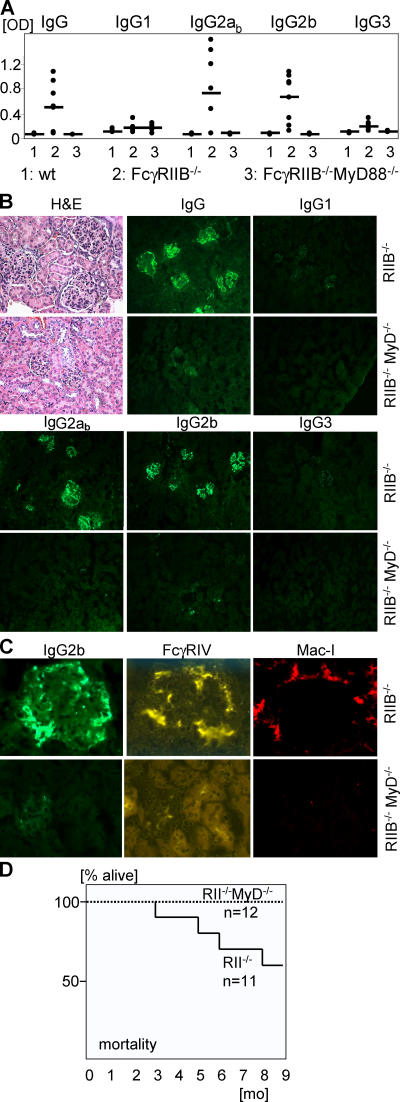
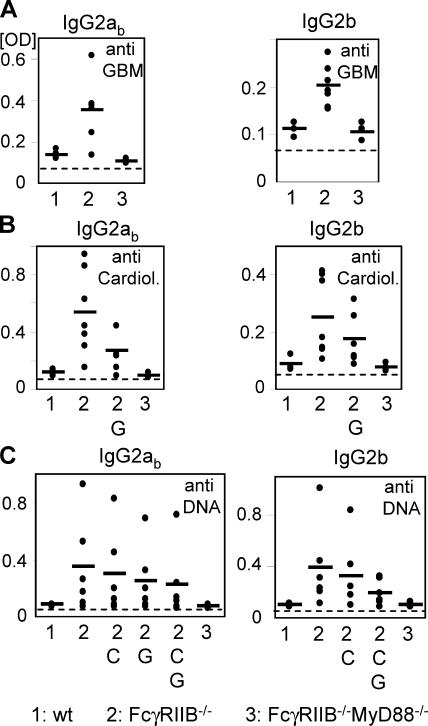
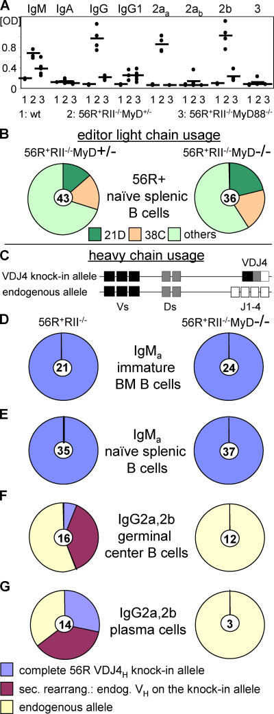
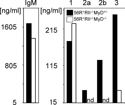
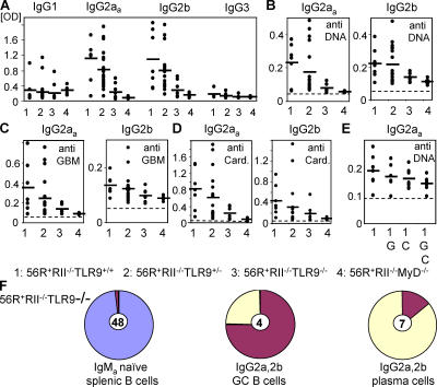
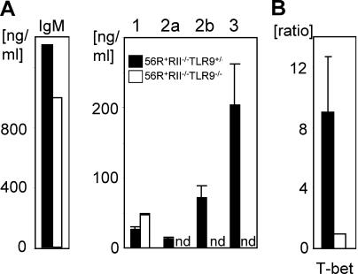
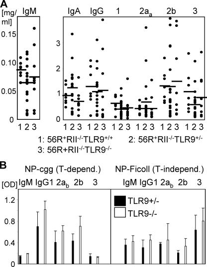
Loss of tolerance in systemic lupus erythematosus (SLE) leads to the generation of autoantibodies, which accumulate in end-organs where they induce disease. Here we show that immunoglobulin (Ig)G2a and 2b autoantibodies are the pathogenic isotypes by recruiting FcgammaRIV expressing macrophages. Class switching, but not development, of IgM anti-self B cells to these pathogenic subclasses requires the innate immune receptor Toll-like receptor (TLR)9 and MyD88 signaling. In their absence, switching of autoreactive B cells to the IgG2a and 2b subclasses is blocked, resulting in reduced pathology and mortality. In contrast, switching of anti-self B cells to IgG1 is not perturbed and generation of nonautoreactive IgG2a and 2b antibodies is not impaired in TLR9-deficient mice. Thus, the TLR9 pathway is a potential target for therapeutic intervention in SLE.


Figures

Figure 1.
Figure 1.
MyD88 deficiency protected against generation and deposition of pathogenic IgG2a and 2b autoantibodies and SLE. (A) IgG subclass analysis of autoantibodies in the serum of 5-mo-old wt.B6 (n = 4), FcγRIIB−/−MyD88+/−.B6 (n = 8), and FcγRIIB−/−MyD88−/−.B6 (n = 6) mice. Shown are anti-DNA and antinuclear antigen titers as determined by ANA ELISA. IgG2ab (haplotype: b) and 2b autoantibodies were up-regulated in FcγRIIB−/−MyD88+/−.B6 mice compared with wt.B6 mice (P = 0.02 and P = 0.004, respectively), whereas IgG2a and 2b titers were reduced to baseline in the absent of MyD88 (P = 0.02 and 0.004, respectively). Horizontal bars represent the average. (B) Histological and immunofluorescence analysis of kidney sections of 9.5-mo-old mice stained with hematoxylin and eosin (H+E) or with anti–mouse IgG subclasses. Compared with the FcγRIIB−/−MyD88+/−.B6 mice, FcγRIIB−/− MyD88−/−.B6 mice showed no IgG2ab and 2b immune complex deposition and pathology in the kidney. (C) Kidney sections in B were costained with anti–mouse IgG2b and anti-FcγRIV (reference 20) or with Mac-I. Sections are representative of three independent mice from each group. (D) Kaplan-Meier survival curves for FcγRIIB−/−MyD88+/+ or FcγRIIB−/−.B6 mice.
Figure 2.
Figure 2.
MyD88 deficiency protected against generation of IgG2a and 2b polyreactive autoantibodies. IgG2ab and 2b analysis of autoantibodies in the serum of 5-mo-old wt.B6 (n = 4), FcγRIIB−/−MyD88+/−.B6 (n = 8), and FcγRIIB−/−MyD88−/−.B6 (n = 6) mice. Shown are antiglomerular basement membrane (GBM) (A), anticardiolipin (Cardiol.) (B), and anti-DNA (C) titers as determined by ELISA. Polyreactivity was analyzed by preabsorbing the samples on the indicated ELISA plates; G: GBM; C: cardiolipin; G,C: GBM + cardiolipin. IgG1 and IgG3 titers were not significantly elevated over baseline (not depicted). Horizontal bars represent the average.
Figure 3.
Figure 3.
IgG2a and IgG2b anti-DNA autoantibodies were absent in the germinal center and plasma cell compartments of MyD88-deficient mice. (A) Isotype and subclass analysis of anti-DNA autoantibodies in the serum of 7–11-wk-old wt.B6 (n = 3), 56R+RIIB−/−MyD88+/− (n = 4), and 56R+RIIB−/−MyD88−/−.B6 (n = 5) mice as determined by ANA ELISA. IgG2aa (haplotype: a) and 2b autoantibodies were strongly and IgG1 slightly up-regulated in 56R+FcγRIIB−/−MyD88+/−.B6 mice compared with wt.B6 mice (P = 0.002, P = 0.004, and P = 0.02, respectively). IgG2ab autoantibodies of B cells without 56R+ were only slightly up-regulated until 7–11 wk in 56R+FcγRIIB−/−MyD88+/−.B6 mice. Among the autoreactive IgG subclasses, IgG1 was unaffected by the absence of MyD88, whereas IgG2a and 2b titers were reduced to baseline (P = 0.002 and 0.03, respectively). Horizontal bars represent the average. (B) MyD-deficient and -sufficient naive splenic B cells used the same light chains, resulting in retention of DNA binding by the 56R VDJ4 heavy chain. Single IgMa + splenic B cells were sorted and single cell PCR was used to identify 56R VH positive cells (knockin allele haplotype: IgMa; C57BL/6 allele haplotype: IgMb). These cells were further analyzed for 21D and 38C κ light chain usage by single cell PCR (reference 6). Naive 56R B cells using the 21D light chain show no DNA binding, whereas 56R B cells using the 38C light chain can still bind DNA (reference 6). (C) Schematic presentation of the anti-DNA 56R knockin VDJ4H transgene. The 56R knockin VDJ4H has replaced the endogenous Js, can be secondarily rearranged with other endogenous Vs, and can class switch to all Ig isotypes (references 22, 23). All 56R+ mice had only one knockin allele. (D and E) MyD88-sufficient (56R+RIIB−/−MyD88+/−) and -deficient IgMa + immature bone marrow (BM) and naive splenic B cells retained the intact 56R VDJ4H transgene. Single IgMa + cells were sorted and analyzed by single cell PCR using a random VH forward primer (reference 34) and an IgM constant region reverse primer (reference 34). (F and G) IgG2a and 2b positive MyD88-deficient GC and splenic plasma cells used the endogenous allele, whereas a fraction of the MyD88-sufficient mice (56R+RIIB−/−MyD88+/− and +/+) still expressed the intact 56R VDJ4H transgene. Single GL7+FAS+ GC or CD138high plasma cells were sorted and analyzed by single cell PCR using a random VH forward primer (reference 34) and specific reverse primers for the different IgG subclass constant regions. A fraction of 56R+RIIB−/− MyD88−/− IgG1 positive GC and plasma cells retained the intact 56R VDJ4 heavy chain (not depicted). No IgG3 cells were found in the GC compartment and no 56R+RIIB−/−MyD88+/− or −/− IgG3 positive plasma cells showed the intact 56R VDJ4 heavy chain (not depicted). The number of cells analyzed is indicated in the center of each pie graph.
Figure 4.
Figure 4.
Loss of IgG2a and 2b autoantibodies in MyD88-deficient mice was B cell intrinsic. Naive splenic B cells from the indicated genotypes were stimulated with 15 μg/ml CpG1826, 10 μg/ml anti-CD40, and 10 ng/ml rmIL-4 in vitro for 5 d and total Ig subclass concentrations in the supernatants were determined. Class switching to IgM was unchanged and to IgG1 increased in MyD88-deficient backgrounds. In contrast, switching to IgG2a, 2b, and 3 was significantly impaired.
Figure 5.
Figure 5.
The TLR9 pathway was required for generation of IgG2a and 2b anti-DNA/polyreactive autoantibodies. IgG subclass analysis of autoantibodies in the serum of 7–11-wk-old 56R+RIIB−/−TLR9+/+.B6 (n = 7), 56R+RIIB−/−TLR9+/−.B6 (n = 19), 56R+RIIB−/−TLR9−/−.B6 (n = 7), or 56R+RIIB−/−MyD88−/−.B6 (n = 6) mice as determined by ANA (A), anti-DNA (B), antiglomerular basement membrane (anti-GBM) (C), and anticardiolipin (D) ELISA, indicated a reduction of the autoreactive IgG2aa and IgG2b subclasses for both genotypes (P = 0.007 and P = 0.03, respectively, for the ANA ELISA). (E) Polyreactivity was analyzed by preabsorbing the samples on the indicated ELISA plates; G: GBM; C: cardiolipin; G,C: GBM + cardiolipin. (F) TLR9-deficient IgMa + naive splenic B cells retained the intact 56R VDJ4H transgene, whereas IgG2a and 2b positive TLR9-deficient GC and splenic plasma cells did not use the intact 56R VDJ4H transgene (compare with Fig. 3).
Figure 6.
Figure 6.
Loss of IgG2a and 2b autoantibodies in TLR9-deficient mice was B cell autonomous. (A) Naive splenic B cells from the indicated genotypes were stimulated with 15 μg/ml CpG1826, 10 μg/ml anti-CD40, and 10 ng/ml rmIL-4 in vitro for 5 d and total Ig subclass concentrations in the supernatants were determined. Class switching to IgM was unchanged and to IgG1 increased in TLR9-deficient backgrounds. In contrast, switching to IgG2a, 2b, and 3 was significantly impaired. (B) T-bet mRNA is not induced by CpG stimulation of B cells in the TLR9-deficient background. A portion of the cells in (A) were collected after 12 h of the indicated stimulation. mRNA and cDNA were prepared and used for real-time PCR with specific primers for T-bet and β-actin as an internal control. The ratio between TLR9-sufficient and -deficient B cells for T-bet mRNA was calculated after normalizing for β-actin. nd, not detected. Results from three independent experiments are shown as mean ± SD.
Figure 7.
Figure 7.
Loss of IgG2a and 2b autoantibodies in TLR9-deficient mice was restricted to anti-self antibodies. (A) Analysis of total antibody isotypes and subclasses in the serum of 7–11-wk-old 56R+RIIB−/− TLR9+/+.B6 (n = 7), 56R+RIIB−/−TLR9+/−.B6 (n = 19), or 56R+RIIB−/− TLR9−/−.B6 (n = 7) mice as determined by ELISA. Horizontal bars represent the average. (B) The specific IgG subclass responses to NP(24)-CGG or NP(24)-Ficoll immunization were not reduced in TLR9−/−.B6 mice compared with TLR9+/−.B6 mice. The indicated anti-NP IgM and IgG subclasses were analyzed on day 11 after injection. Results from three independent experiments are shown as mean ± SD.

References

    1. Peter, J.B., and Y. Shoenfeld. 1996. Autoantibodies. Elsevier, New York. 910 pp.
    1. Bolland, S., and J.V. Ravetch. 2000. Spontaneous autoimmune disease in FcγRII deficient mice results from strain-specific epistasis. Immunity. 13:277–285. - PubMed
    1. Rifkin, I.R., and A. Marshak-Rothstein. 2003. T-bet: the Toll-bridge to class-switch recombination? Nat. Immunol. 4:650–652. - PubMed
    1. Fields, M.L., and J. Erikson. 2003. The regulation of lupus-associated autoantibodies: immunoglobulin transgenic models. Curr. Opin. Immunol. 15:709–717. - PubMed
    1. Wakeland, E.K., K. Liu, R.R. Graham, and T.W. Behrens. 2001. Delineating the genetic basis of systemic lupus erythematosus. Immunity. 15:397–408. - PubMed

Publication types

MeSH terms