Comparative Study
. 2006 Feb 27;172(5):679-92.
doi: 10.1083/jcb.200508154. Epub 2006 Feb 21.

Repo-Man recruits PP1 gamma to chromatin and is essential for cell viability

Affiliations
Comparative Study

Repo-Man recruits PP1 gamma to chromatin and is essential for cell viability

Laura Trinkle-Mulcahy et al. J Cell Biol. .

Abstract

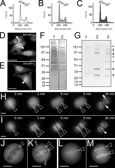
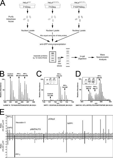
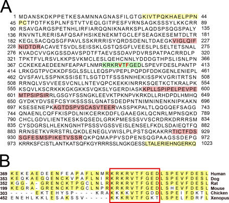
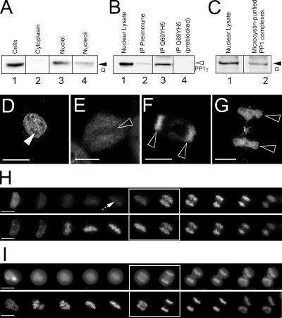
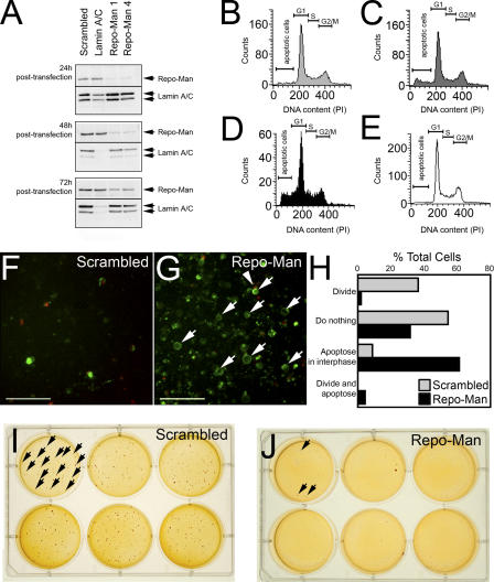
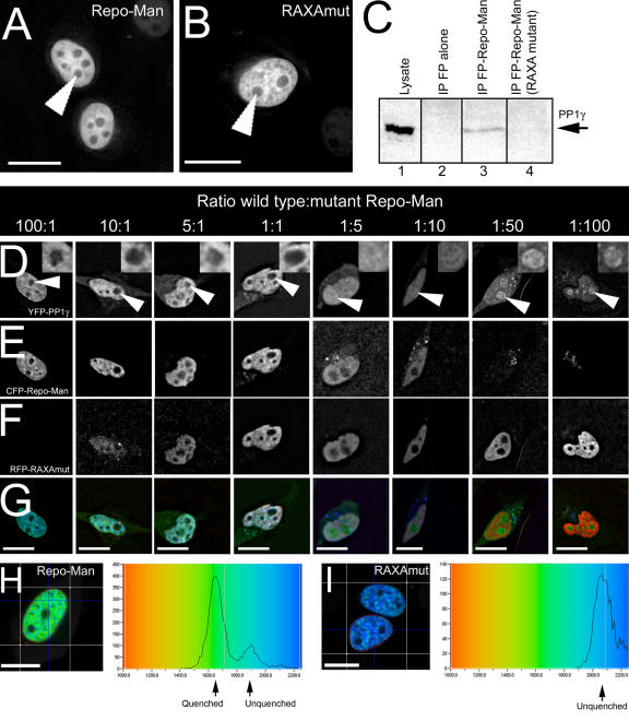
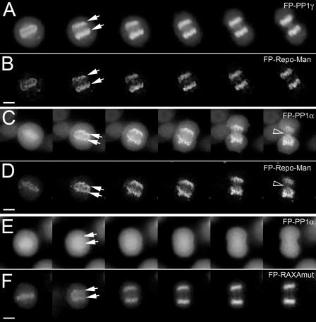
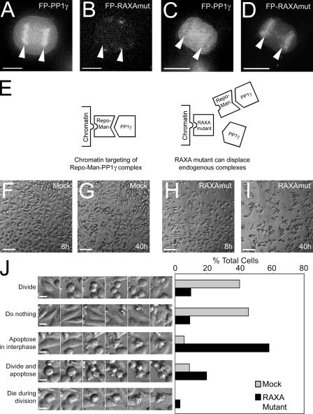
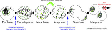
Protein phosphatase 1 (PP1) is a ubiquitous serine/threonine phosphatase regulating many cellular processes. PP1alpha and -gamma are closely related isoforms with distinct localization patterns, shown here by time-lapse microscopy of stably expressed fluorescent protein fusions. A pool of PP1gamma is selectively loaded onto chromatin at anaphase. Using stable isotope labeling and proteomics, we identified a novel PP1 binding protein, Repo-Man, which selectively recruits PP1gamma onto mitotic chromatin at anaphase and into the following interphase. This approach revealed both novel and known PP1 binding proteins, quantitating their relative distribution between PP1alpha and -gamma in vivo. When overexpressed, Repo-Man can also recruit PP1alpha to chromatin. Mutating Repo-Man's PP1 binding domain does not disrupt chromatin binding but abolishes recruitment of PP1 onto chromatin. RNA interference-induced knockdown of Repo-Man caused large-scale cell death by apoptosis, as did overexpression of this dominant-negative mutant. The data indicate that Repo-Man forms an essential complex with PP1gamma and is required for the recruitment of PP1 to chromatin.


Figures

Figure 1.
Figure 1.
Intrinsic differences between FP-PP1α and FP-PP1γ expressed stably in mammalian cells. (A–C) FACS analyses show no significant difference in cell cycle distribution between HeLaEGFP-PP1α (B) and HeLaEGFP-PP1γ (C) cells and parental HeLa cells (A). (D and E) Fluorescence imaging of the stable cell lines during interphase reveals distinct localization patterns for FP-PP1α (D) and FP-PP1γ (E), with nucleoli (arrowheads) and FP-PP1α foci (arrow) marked. (F) FP-PP1α (lane 1) and FP-PP1γ (lane 2) immunoprecipitates separated by one-dimensional SDS-PAGE and silver stained. (G) Far Western overlay with 35S-PP1 shows differences (arrowheads) between FP-PP1α (lane 2) and FP-PP1γ (lane 3) immunoprecipitates. FP alone (lane 1) does not coprecipitate PP1 binding proteins. (H and I) Time-lapse imaging of HeLaEGFP-PP1α (H) and HeLaEGFP-PP1γ (I) cells during mitosis shows differences in localization with respect to chromatin (open arrowheads). Although both accumulate at kinetochores in metaphase (0 min), FP-PP1α is excluded from other chromatin regions, reaccumulating within the nucleus and nuclear foci (solid arrow) in late telophase (36 min), with additional accumulations at the midbody and cell cortex (dotted arrow). In contrast, a large pool of FP-PP1γ appears on chromatin at anaphase (3 min) and remains there, with a pool reentering nucleoli in late telophase (closed arrowhead). (J–M) High-resolution imaging of live mitotic HeLaEGFP-PP1α cells shows the kinetochore localization and chromatin (arrowheads) exclusion of FP-PP1α during metaphase (J) and anaphase (K), as compared with FP-PP1γ's kinetochore localization at metaphase (L) and sudden chromatin association at anaphase (M). A pool of FP-PP1α is also found on centrosomes (arrows). Bars, 10 μm.
Figure 2.
Figure 2.
Identification of PP1 binding proteins by combined SILAC/mass spectrometry. (A) Design of the SILAC experiment. (B–D) Sample peptide spectra measured, illustrating the shift in peptide mass caused by incorporation of heavy arginine isotopes. Proteins that copurify with FP, FP-PP1α, and FP-PP1γ show a 1:1:1 ratio (B). Proteins showing specific interactions with either FP-PP1α (C) or FP-PP1γ (D) reflect that in their arginine ratios, which are in agreement with those calculated by quantitation of protein levels via Western blotting (C and D, insets). (E) Proteins identified in one experiment, listed in order of database identification score and plotted as relative abundance ratios (FP-PP1α/FP plotted as positive values and FP-PP1γ/FP plotted as negative values). Ratios >1.5 are taken to indicate specific coimmunoprecipitation with FP-PP1. PP1α and -γ peptides were also found, with the expected arginine ratios for their respective cell lines.
Figure 3.
Figure 3.
Sequence analysis of Q69YH5. (A) Amino acid sequence for Q69YH5, highlighting peptides identified by mass spectrometry (pink), peptides used to raise affinity-purified antibodies (yellow), and the putative RVXF (PP1 binding) motif (green). For mutational analysis, the hydrophobic Val and Phe residues within this region (red) were mutated to Ala. (B) Clustal alignment of this region in Q69YH5 protein sequences from several vertebrates. Identical residues are highlighted in yellow, and the highly conserved RVXF motif is outlined in red. The full sequence alignment is shown in Fig. S1, available at http://www.jcb.org/cgi/content/full/jcb.200511068/DC1.
Figure 4.
Figure 4.
Q69YH5 interacts with endogenous PP1 and localizes to chromatin. (A) Western blot of HeLa cell fractions with anti-Q69YH5 peptide antibodies shows that Q69YH5 is a predominantly nuclear protein (lane 3, arrowhead). (B) When immunoprecipitated from interphase nuclear lysates, endogenous Q69YH5 coprecipitates endogenous PP1γ (lane 3, arrowhead). PP1 does not coprecipitate significantly with preimmune sera (lane 2) or with anti-Q69YH5 antibody pretreated with the cognate peptide (lane 4). (C) Purification of endogenous PP1 complexes from interphase nuclear lysates using microcystin affinity chromatography also shows that endogenous Q69YH5 (lane 2, arrowhead) is in a complex with PP1. (D–G) Staining of endogenous Q69YH5 with anti-Q69HY5 antibody reveals that is predominantly nucleoplasmic and excluded from nucleoli (closed arrowhead) during interphase (D). Q69YH5 is predominantly diffuse in metaphase cells (E) but shows an accumulation on chromatin (open arrowheads) during anaphase (F) and telophase (G). (H) This dynamic localization was confirmed by time-lapse imaging of HeLa cells transiently expressing FP-Q69YH5 (top) stained with Hoechst 33342 (bottom). Images were taken every 3 min as the cell progressed from prophase to telophase. There is a faint accumulation of FP-Q69YH5 on chromatin at metaphase (arrow). (I) For comparison, a similar time-lapse profile is shown for a HeLaEGFP-PP1γ cell (top) stained with Hoechst 33342 (bottom). The white boxes indicate the time at which a significant recruitment of both FP-Q69YH5 and FP-PP1γ onto chromatin is observed. Bars, 10 μm.
Figure 5.
Figure 5.
Depletion of Repo-Man by RNAi induces apoptosis and decreases cell viability. (A) Western blot analyses of HeLa cells treated with various siRNA duplexes. Lysates were prepared 24, 48, and 72 h after transfection. (B–D) FACS profiles for cells treated with Repo-Man duplex 1 and fixed at 24 h (B), 48 h (C), and 72 h (D) after transfection. (E) For comparison, a FACS profile for cells treated with scrambled duplex and fixed at 72 h is shown. (F and G) Cells treated with either scrambled duplex (F) or Repo-Man duplex 1 (G) for 24 h were also stained with Annexin V–FITC (green, arrows) and propidium iodide (red, arrowheads) to detect cells in early stages of apoptosis. Bars, 40 μm. (H) Graph summarizes the fates observed for HeLa cells by DIC imaging 24–48 h after transfection with either scrambled duplex (gray bars; n = 90) or Repo-Man duplex 1 (black bars; n = 88). The graph shows the number of cells in each category as a percentage of total cells. (I and J) Soft agar assays were performed to detect cell viability via colony formation (arrows) 2 wk after transfection with either scrambled duplex (I) or Repo-Man duplex 1 (J).
Figure 6.
Figure 6.
Mutation of the RVXF motif in Repo-Man disrupts interaction with PP1. (A and B) When transiently expressed, RAXA mutant FP–Repo-Man (B) shows a similar nucleoplasmic accumulation at interphase to wild type (A). Nucleoli are marked by arrowheads. (C) Unlike wild-type FP–Repo-Man (lane 3), the RAXA mutant does not coprecipitate endogenous PP1γ (lane 4), as shown by Western blotting. (D–G) Transient expression of wild-type but not RAXA mutant FP–Repo-Man also retargets exogenous PP1, as demonstrated by titrating varying levels of the two plasmids into HeLaEYFP-PP1γ cells. FP-PP1γ is shown in D (nucleoli indicated by arrowheads are enlarged in inset to compare PP1 levels), wild-type FP–Repo-Man in E, and RAXA mutant FP–Repo-Man in F. Merged images are shown in G. (H and I) A direct interaction is measured between wild-type CFP–Repo-Man and YFP-PP1γ using fluorescence lifetime imaging microscopy/FRET analysis (H) but not between RAXA mutant CFP–Repo-Man and YPF-PP1γ (I). Color-coded fluorescence lifetime maps are shown for each CFP construct (donor) in the presence of YFP-PP1γ (acceptor) and the respective quenched and unquenched lifetimes indicated on the graphs. Bars, 10 μm.
Figure 7.
Figure 7.
Overexpression of FP–Repo-Man ectopically recruits FP-PP1α to chromatin. (A and B) A series of time-lapse images taken every 3 min of a HeLaEYFP-PP1γ cell (A) transiently expressing ECFP–Repo-Man (B) as it progresses from metaphase to telophase. Arrows mark chromatin regions. (C and D) Time-lapse series for a HeLaEGFP-PP1α cell transiently expressing monomeric dsRed–Repo-Man (D), which retargets a significant pool of FP-PP1α (C) to chromatin at anaphase (arrows) and retains it there. Note the lagging chromosomes observed under these conditions (arrowheads). (E and F) Expression of RAXA mutant FP–Repo-Man (F) does not cause this retargeting of FP-PP1α (E). Bars, 10 μm.
Figure 8.
Figure 8.
Overexpression of RAXA mutant FP–Repo-Man displaces endogenous Repo-Man–PP1γ complexes from chromatin. (A–D) When RAXA mutant FP–Repo-Man (D) is expressed in HeLaEGFP-PP1γ cells, significantly lower levels of FP-PP1γ (C) are found on anaphase chromatin (arrowheads), compared with the levels observed (A) in cells expressing little or no mutant FP–Repo-Man (B). Bars, 10 μm. This indicates a dominant-negative effect, as depicted by E. (F–I) Time-lapse DIC imaging of HeLa cells after mock-transfection (F and G) or transfection with RAXA mutant FP–Repo-Man (H and I) was used to quantify the extent and type of cell death caused by these conditions, between 8 h (F and H) and 40 h (G and I) after transfection. Bars, 100 μm. (J) Summary of the fates observed for HeLa cells by DIC imaging after either mock-transfection (gray bars; n = 90) or transfection with RAXA mutant monomeric dsRed–Repo-Man (black bars; n = 163). Cell fates were divided into five categories as indicated, and the graph shows the number of cells in each category as a percentage of total cells. Bars, 10 μm.
Figure 9.
Figure 9.
Localization of Repo-Man-PP1γ complexes throughout the cell cycle. Based on the data presented in this study, we provide a depiction of the localization of Repo-Man-PP1γ complexes (green) throughout the cell cycle, noting its initial loading onto chromatin at the metaphase-anaphase transition. Also shown is the key stage (red) at which both RNAi knockdown of endogenous Repo-Man and displacement of endogenous Repo-Man-PP1 complexes by the dominant-negative mutant induce apoptosis in interphase cells. Observed effects of overexpression of wild-type Repo-Man on mitotic cells are also indicated (blue).

Similar articles

Cited by

References

    1. Allen, P.B., Y.G. Kwon, A.C. Nairn, and P. Greengard. 1998. Isolation and characterization of PNUTS, a putative protein phosphatase 1 nuclear targeting subunit. J. Biol. Chem. 273:4089–4095. - PubMed
    1. Andersen, J.S., C.E. Lyon, A.H. Fox, A.K. Leung, Y.W. Lam, H. Steen, M. Mann, and A.I. Lamond. 2002. Directed proteomic analysis of the human nucleolus. Curr. Biol. 12:1–11. - PubMed
    1. Andersen, J.S., Y.W. Lam, A.K. Leung, S.E. Ong, C.E. Lyon, A.I. Lamond, and M. Mann. 2005. Nucleolar proteome dynamics. Nature. 433:77–83. - PubMed
    1. Andreassen, P.R., F.B. Lacroix, E. Villa-Moruzzi, and R.L. Margolis. 1998. Differential subcellular localization of protein phosphatase-1 α, γ1, and δ isoforms during both interphase and mitosis in mammalian cells. J. Cell Biol. 141:1207–1215. - PMC - PubMed
    1. Blagoev, B., S.E. Ong, I. Kratchmarova, and M. Mann. 2004. Temporal analysis of phosphotyrosine-dependent signaling networks by quantitative proteomics. Nat. Biotechnol. 22:1139–1145. - PubMed

Publication types